. 2005 Oct;115(10):2904-13.
doi: 10.1172/JCI23961. Epub 2005 Sep 15.

TCR stimulation with modified anti-CD3 mAb expands CD8+ T cell population and induces CD8+CD25+ Tregs

Affiliations

TCR stimulation with modified anti-CD3 mAb expands CD8+ T cell population and induces CD8+CD25+ Tregs

Brygida Bisikirska et al. J Clin Invest. 2005 Oct.

Abstract

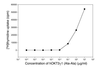
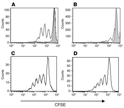
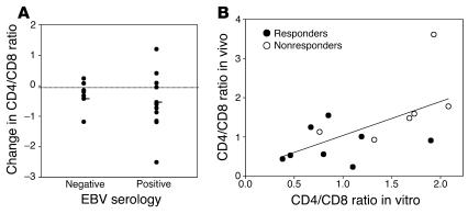
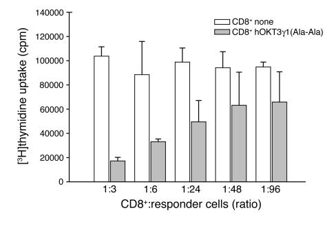
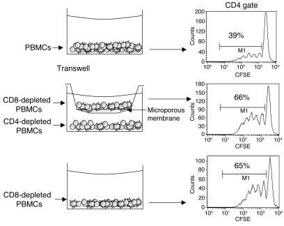
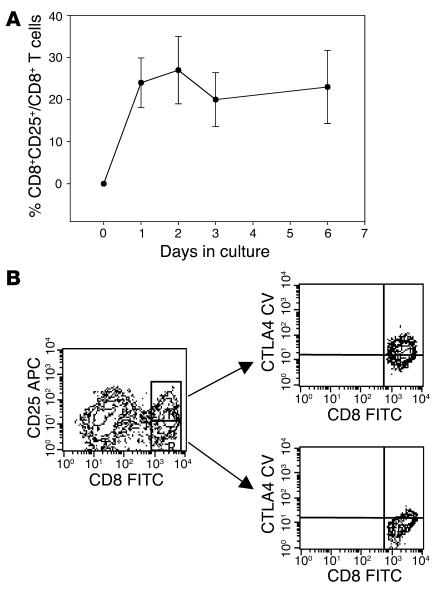
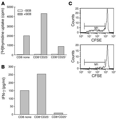
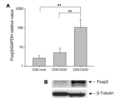
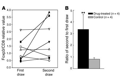
Modified anti-CD3 mAbs are emerging as a possible means of inducing immunologic tolerance in settings including transplantation and autoimmunity such as in type 1 diabetes. In a trial of a modified anti-CD3 mAb [hOKT3gamma1(Ala-Ala)] in patients with type 1 diabetes, we identified clinical responders by an increase in the number of peripheral blood CD8+ cells following treatment with the mAb. Here we show that the anti-CD3 mAb caused activation of CD8+ T cells that was similar in vitro and in vivo and induced regulatory CD8+CD25+ T cells. These cells inhibited the responses of CD4+ cells to the mAb itself and to antigen. The regulatory CD8+CD25+ cells were CTLA4 and Foxp3 and required contact for inhibition. Foxp3 was also induced on CD8+ T cells in patients during mAb treatment, which suggests a potential mechanism of the anti-CD3 mAb immune modulatory effects involving induction of a subset of regulatory CD8+ T cells.


Figures

Figure 1
Figure 1
Anti-CD3 hOKT3γ1(Ala-Ala) antibody stimulates in vitro proliferation of human PBMCs. PBMCs were cultured for 5 days in the presence of hOKT3γ1(Ala-Ala) antibody at the indicated concentrations. Cell proliferation level was determined by [3H]thymidine uptake. Results are expressed as the mean value of duplicates and are representative of 3 independent experiments.
Figure 2
Figure 2
Diverse in vitro response of CD8+ and CD4+ subpopulations of human PBMCs to hOKT3γ1(Ala-Ala) stimulation. CFSE-labeled PBMCs were cultured in the presence of hOKT3γ1(Ala-Ala) (A and B) or PHA (C and D) for 6 days. Cells were labeled with fluorochrome-conjugated anti-CD4 and anti-CD8 and analyzed by flow cytometry. (A and C) CD8-gated cells. (B and D) CD4-gated cells. Gray histograms: cells without stimulation. Results from a single experiment representative of 6 experiments are shown.
Figure 3
Figure 3
Changes in CD4/CD8 T cell ratio in subjects with type 1 diabetes receiving hOKT3γ1(Ala-Ala) in relation to EBV status at study entry, and correlation between changes in CD4+ and CD8+ T cells in vitro during culture with anti-CD3 mAb and in vivo following treatment with anti-CD3 mAb. (A) The difference in the CD4/CD8 T cell ratio 3 months after treatment with hOKT3γ1(Ala-Ala) from the ratio before drug treatment in individuals who were EBV seropositive (n = 12) or seronegative (n = 7) at study entry was determined. The dark lines indicate the mean values for the group. A decrease in the CD4/CD8 T cell ratio (below the dotted line) occurred in both EBV-seropositive and -seronegative subjects. (B) PBMCs from patients with type 1 diabetes mellitus who received anti-CD3 mAb were studied 1.5–2 years after mAb treatment, at which time the changes in CD4/CD8 T cell ratio seen after mAb treatment had resolved. The patients were designated as clinical responders (filled circles) or nonresponders (open circles) based on their C-peptide responses at 12 months compared with baseline. The cells were cultured with hOKT3γ1(Ala-Ala), and the percentages of CD4+ and CD8+ T cells were determined after 6 days. The Pearson correlation coefficient was calculated to compare the ratio of CD4+/CD8+ T cells in vitro with the analysis of CD4+/CD8+ T cells in vivo 3 months after mAb treatment (8), and the level of significance was tested with correlation procedure SAS (r = 0.6; P = 0.024). The line indicates the relationship between the changes in vitro and in vivo [in vivo CD4/CD8 ratio = 0.174 + 0.865 (in vitro CD4/CD8 ratio)].
Figure 4
Figure 4
Reduced proliferative response of CD4+ cells to hOKT3γ1(Ala-Ala) stimulation occurs only in the presence of CD8+ cells but is not due to lack of IL-2. CFSE-labeled cells were cultured in the presence of hOKT3γ1(Ala-Ala) for 6 days. Cells were stained with PE-conjugated anti-CD4 and anti-CD25 mAbs and analyzed on a FACSCalibur flow cytometer. (A and C) Bulk PBMCs. (B and D) PBMCs depleted of CD8+ T cells. Representative results of 6 independent experiments are shown. The numbers over the dots in A and B represent the number of cell divisions. The percentages in C and D represent the percentage of CD4+ cells that were CD25+. PBMCs were cultured with the anti-CD3 mAb in the absence (E and F) or presence (G and H) of recombinant IL-2 (50 U/ml). The addition of IL-2 to the cultures enhanced proliferation of CD8+ T cells (E and G) but did not have an effect on CD4+ T cell proliferation in the PBMCs (F and H). Representative results of 4 independent experiments are shown.
Figure 5
Figure 5
CD8+ lymphocytes from PBMC cultures stimulated with hOKT3γ1(Ala-Ala) suppress tetanus-specific response. CD8 cells isolated from fresh PBMCs (CD8 none; white bars) or from PBMCs cultured for 6 days in the presence of hOKT3γ1(Ala-Ala) (gray bars) were irradiated and mixed with fresh PBMCs depleted of CD8+ T cells at the indicated ratios. Cells were cultured with or without the presence of tetanus toxoid for 3 days. Cell proliferation was determined by [3H]thymidine uptake, and the data are expressed as the difference (in counts per minute) between cultures with and without antigen. The background counts (responder cells with CD8+ cells in the absence of antigen) ranged between 542 and 1,796 cpm. Representative results of 3 independent experiments are shown.
Figure 6
Figure 6
Suppression of CD4+ cell proliferation by CD8+ lymphocytes is not mediated by soluble factors. CFSE-labeled cells were cultured in the presence of hOKT3γ1(Ala-Ala) for 6 days. Cells were labeled with fluorochrome-conjugated anti-surface marker mAb and analyzed by flow cytometry. Histograms were gated on CD4+ lymphocytes. Shown are PBMCs (top panel), CD8-depleted PBMCs separated by Transwell membrane from CD4-depleted PBMCs (middle panel), and CD8-depleted PBMCs (bottom panel). The numbers in each histogram represent the percentage of CD4+ cells with dilution of CFSE. Results are representative of 4 independent experiments.
Figure 7
Figure 7
Induction of CD25+ population in CD8 cells stimulated with hOKT3γ1(Ala-Ala). (A) Freshly isolated PBMCs were cultured in the presence of hOKT3γ1(Ala-Ala) for 6 days. For analysis of CD25 expression, cells were collected on days 0 (before stimulation), 1, 2, 3, and 6 of the culture, labeled with flourochrome-conjugated anti-CD8 and anti-CD25, and analyzed by flow cytometry. The results are presented as a ratio of CD8+CD25+-expressing cells to total CD8+ cells. Mean values (± SEM) of 4 independent experiments are shown. (B) Intracellular expression of CTLA4 analyzed by flow cytometry. Left: gated CD8 lymphocytes; upper right: gated CD8+CD25+ cells; lower right: gated CD8+CD25 cells. Representative results from 3 independent experiments are shown. CTLA4CV, CTLA4 CyChrome.
Figure 8
Figure 8
The CD8+CD25+ cells induced with hOKT3γ1(Ala-Ala) suppress CD4 cell response to SEB and IFN-γ secretion. (A) CD8+CD25+ or CD8+CD25 cells sorted from PBMCs stimulated for 6 days with hOKT3γ1(Ala-Ala) or CD8+ cells from fresh PBMCs were irradiated and cocultured for 3 days with fresh PBMCs depleted of CD8 in the presence of SEB. Proliferative response was assessed by [3H]thymidine uptake. (B) The levels of IFN-γ in the supernatants from the same cultures were measured by Luminex system using Th1/Th2 multiplex microspheres. Representative results of 2 independent experiments are shown. (C) Sorted CD8+CD25+ (bottom panel) or untreated CD8 cells (upper panel) were cocultured for 6 days with CFSE-labeled, CD8-depleted PBMCs at 1:2 ratio in the presence of SEB. SEB-specific clonal expansion was analyzed by flow cytometry. The expansion of Vβ3-positive CD4 (M1) lymphocytes was reduced from 46.1% to 15.5% of Vβ3+ T cells. Similar results were obtained in 2 additional studies.
Figure 9
Figure 9
Increased expression of Foxp3 in CD8+CD25+ cells induced with hOKT3γ1(Ala-Ala). (A) PBMCs were cultured for 6 days with hOKT3γ1(Ala-Ala) and sorted based on the expression of CD25. Foxp3 expression was measured by quantitative real-time PCR. Results are presented as Foxp3 gene expression normalized to GAPDH expression, and the results for CD8+CD25+ or CD8+CD25 cells were compared with those for freshly isolated CD8+ T cells from the same subject. Mean values (±SD) of 4 independent experiments are shown. **P ≤ 0.02. (B) Expression of Foxp3 was studied in CD8+CD25+ and CD8+CD25 cells from the same cultures by Western blotting and was detected at higher levels in CD8+CD25+ cells.
Figure 10
Figure 10
Changes in Foxp3 expression in vivo in CD8+ PBMCs following treatment with anti-CD3 mAb. CD8+ T cells were isolated from the same individual before (first draw) and after (second draw) treatment with anti-CD3 mAb (filled symbols; n = 4) or in control subjects with type 1 diabetes (open symbols) on 2 (n = 3) or 3 (n = 1) occasions. The expression of Foxp3 relative to CD8 (×100) for each individual is plotted (A), and the average ratio of the second/first sampling in each group (± SEM) is shown (B). **P = 0.02.

References

    1. Xu D, et al. In vitro characterization of five humanized OKT3 effector function variant antibodies. Cell Immunol. 2000;200:16–26. - PubMed
    1. Carpenter PA, Tso JY, Press OW, Yu X, Anasetti C. Non-FcR-binding, humanized anti-CD3 antibody Hu291 induces apoptosis of human T cells more effectively than OKT3 and is immunosuppressive in vivo. Transplant. Proc. 2000;32:1545–1546. - PubMed
    1. Bolt S, et al. The generation of a humanized, non-mitogenic CD3 monoclonal antibody which retains in vitro immunosuppressive properties. Eur. J. Immunol. 1993;23:403–411. - PubMed
    1. Smith JA, Tso JY, Clark MR, Cole MS, Bluestone JA. Nonmitogenic anti-CD3 monoclonal antibodies deliver a partial T cell receptor signal and induce clonal anergy. J. Exp. Med. 1997;185:1413–1422. - PMC - PubMed
    1. Utset TO, et al. Modified anti-CD3 therapy in psoriatic arthritis: a phase I/II clinical trial. J. Rheumatol. 2002;29:1907–1913. - PubMed

Publication types

MeSH terms