Review
. 2005 Jun;37(3):143-53.
doi: 10.1007/s10863-005-6567-7.

Mitochondrial ceramide and the induction of apoptosis

Affiliations
Review

Mitochondrial ceramide and the induction of apoptosis

Leah J Siskind. J Bioenerg Biomembr. 2005 Jun.

Abstract

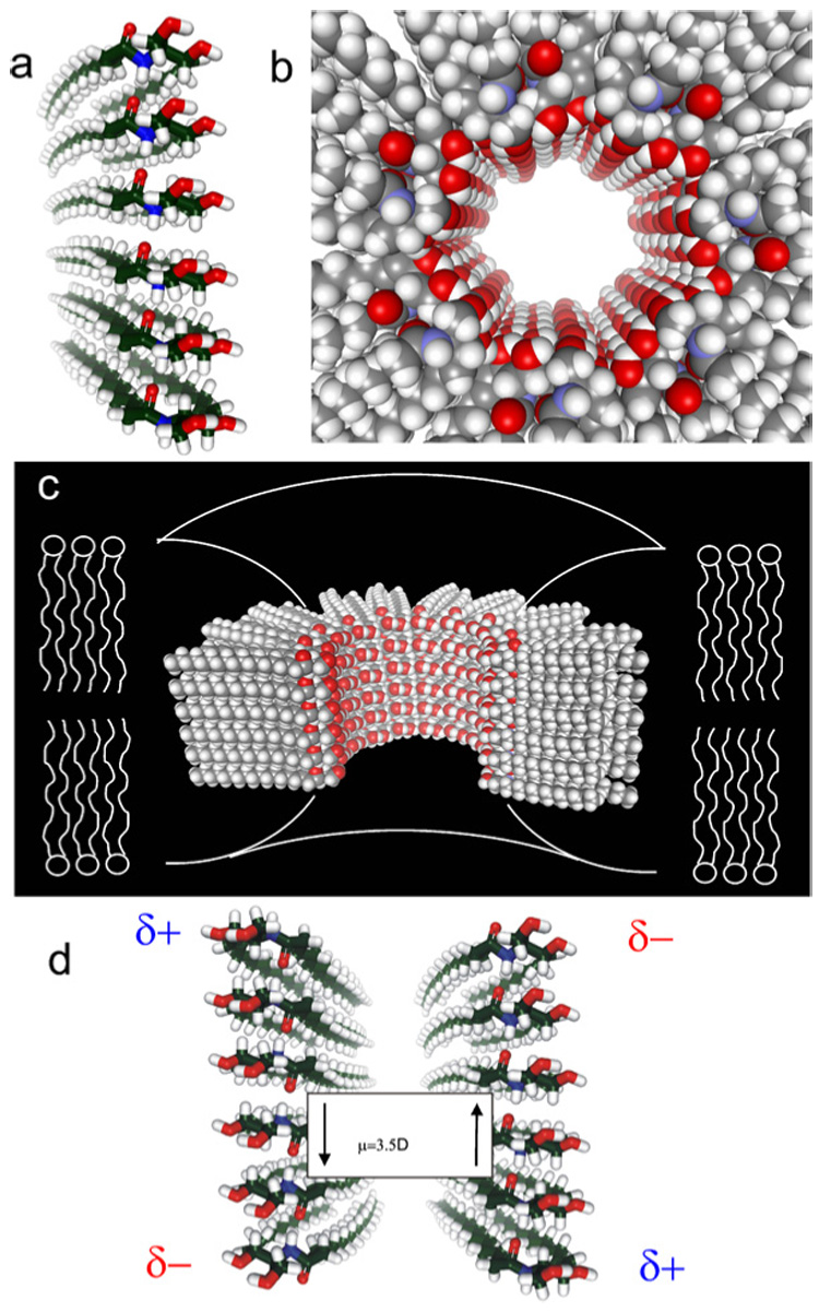
In most cell types, a key event in apoptosis is the release of proapoptotic intermembrane space proteins from mitochondria to the cytoplasm. In general, it is the release of these intermembrane space proteins that is responsible for the activation of caspases and DNases that are responsible for the execution of apoptosis. The mechanism for the increased permeability of the mitochondrial outer membrane during the induction phase of apoptosis is currently unknown and highly debated. This review will focus on one such proposed mechanism, namely, the formation of ceramide channels in the mitochondrial outer membrane. Ceramides are known to play a major regulatory role in apoptosis by inducing the release of proapoptotic proteins from the mitochondria. As mitochondria are known to contain the enzymes responsible for the synthesis and hydrolysis of ceramide, there exists a mechanism for regulating the level of ceramide in mitochondria. In addition, mitochondrial ceramide levels have been shown to be elevated prior to the induction phase of apoptosis. Ceramide has been shown to form large protein permeable channels in planar phospholipid and mitochondrial outer membranes. Thus, ceramide channels are good candidates for the pathway with which proapoptotic proteins are released from mitochondria during the induction phase of apoptosis.


Figures

Fig. 1
Fig. 1
Ceramides form channels in phospholipid membranes. (a) General structure of a ceramide molecule, which consists of a sphingoid base backbone and a N-linked fatty acyl chain that can vary in length from a n = 1 to more than 23. (b) Example ceramide conductance increments observed following the addition of 5 µM C22-ceramide to the aqueous phase on one side of a solvent free planar phospholipid membrane formed from monolayers composed of 0.5% (w/v). DiPyPC, 0.5% (w/v) asolectin, 0.2% (w/v) cholesterol. The applied voltage was clamped at 10 mV (trans side was ground). The aqueous solution bathing both sides of the membrane was composed of 1 M KCl, 1 mM MgCl2, 5 mM PIPES (pH 6.8).
Fig. 2
Fig. 2
Ceramides form protein permeable channels in mitochondrial outer membranes of isolated mitochondria. (a) Ceramide addition to isolated rat liver mitochondrial suspensions allows the oxidation of exogenously added reduced cytochrome c. Mitochondria were incubated with the following treatments: mitochondria controls (M; untreated mitochondria, vehicle controls); 20 µM C2-dihydroceramide for 10 min (DH); 20 µM C2-ceramide for 10 min (C2); 20 µM C16-ceramide for 10 min (C16); mitochondria with lysed outer membranes were incubated with 20 µM C2-dihydroceramide for 10 min (L-DH); lysed mitochondrial controls (L; untreated lysed mitochondria, lysed mitochondria incubated with C2- and C16-ceramide for 10 min, lysed mitochondria vehicle controls). Reduced cytochrome c was then added and its oxidation monitored by measuring the absorbance decrease at 550 nm. (b) The permeability increase induced by C2-ceramide can be reversed with bovine serum albumin (BSA). Isolated mitochondria were incubated with 20 µM C2-ceramide and were indicated 83 µM BSA for the indicated time periods. Reduced cytochrome c was then added and the absorbance was then monitored at 550 nm. (c) C2- and C16-ceramide increase the permeability of the mitochondrial outer membrane to intermembrane space proteins with a cut-off of 60 kDa. Mitochondria were incubated with C2- or C16-ceramides or C2-dihydroceramide at a level of 18 nmol ceramide added/mg mitochondrial protein for 10 min. Released proteins were run on a 15% acrylamide SDS-PAGE and the gel stained with GelCode Blue stain (Pierce). Densitometry from the gel was performed and the individual background for each lane on the gel and the untreated mitochondrial control subtracted out. The results were expressed as a percent of the total protein (mitochondria with lysed outer membranes) versus the RF value.
Fig. 3
Fig. 3
Structural model for ceramide channels. (a) A column of ceramide residues held together by intermolecular hydrogen bonds between amide nitrogens and carbonyl groups. This column would span the hydrophobic portion of the membrane and in association with other columns would form channels of varying sizes. (b) Top view of a ceramide channel consisting of 12 columns of ceramide molecules. Adjacent columns are oriented in an antiparallel fashion so that amide dipoles attract. The columns are held together via intermolecular hydrogen bonds between hydroxyl groups proposed to line the channel lumen. (c) A longitudinal cut-away of a ceramide channel, consisting of 14 columns of ceramide molecules, where 4 columns have been removed to show the interior of the channel. The curvature of the phospholipids of the membrane at the channel interface would minimize the exposure of the hydrophobic regions of the outer surface of the channel to the aqueous solution. (d) Two adjacent ceramide columns oriented in an antiparallel fashion so that opposite dipoles attract. This antiparallel orientation adds to the stability of the channel.

References

    1. Allouche M, Bettaieb A, Vindis C, Rousse A, Grignon C, Laurent G. Oncogene. 1997;14:1837–1845. - PubMed
    1. Alphonse G, Aloy MT, Broquet P, Gerard JP, Louisot P, Rousson R, Rodriguez-Lafrasse C. Int. J. Radiat. Biol. 2002;78:821–835. - PubMed
    1. Alphonse G, Bionda C, Aloy M-T, Ardail D, Rousson R, Rodriguez-Lafrasse C. Oncogene. 2004;23:2703–2715. - PubMed
    1. Amarante-Mendes GP, Naekyung Kim C, Liu L, Huang Y, Perkins CL, Green DR, Bhalla K. Blood. 1998;91:1700–1705. - PubMed
    1. Ardail D, Popa I, Alcantara K, Pons A, Zanetta JP, Louisot P, Thomas L, Portoukalian J. FEBS Lett. 2001;488:160–164. - PubMed

Publication types