Review
. 2005;9 Suppl 4(Suppl 4):S13-9.
doi: 10.1186/cc3753. Epub 2005 Aug 25.

The microcirculation is the motor of sepsis

Affiliations
Review

The microcirculation is the motor of sepsis

Can Ince. Crit Care. 2005.

Abstract

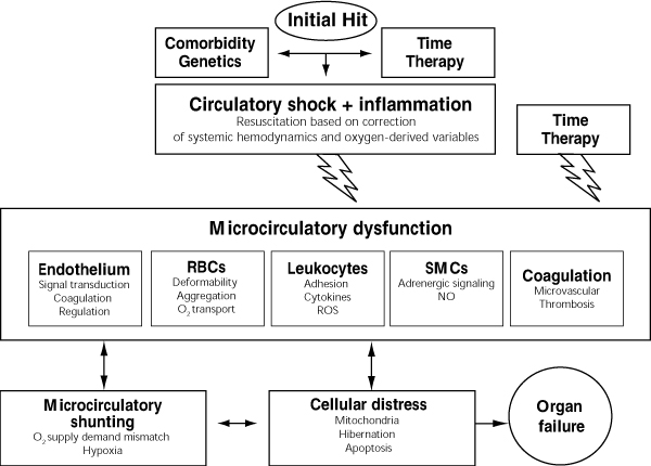
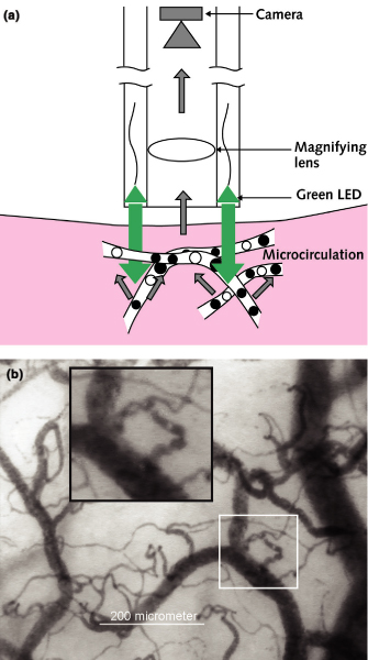
Regional tissue distress caused by microcirculatory dysfunction and mitochondrial depression underlies the condition in sepsis and shock where, despite correction of systemic oxygen delivery variables, regional hypoxia and oxygen extraction deficit persist. We have termed this condition microcirculatory and mitochondrial distress syndrome (MMDS). Orthogonal polarization spectral imaging allowed the first clinical observation of the microcirculation in human internal organs, and has identified the pivotal role of microcirculatory abnormalities in defining the severity of sepsis, a condition not revealed by systemic hemodynamic or oxygen-derived variables. Recently, sublingual sidestream dark-field (SDF) imaging has been introduced, allowing observation of the microcirculation in even greater detail. Microcirculatory recruitment is needed to ensure adequate microcirculatory perfusion and the oxygenation of tissue cells that follows. In sepsis, where inflammation-induced autoregulatory dysfunction persists and oxygen need is not matched by supply, the microcirculation can be recruited by reducing pathological shunting, promoting microcirculatory perfusion, supporting pump function, and controlling hemorheology and coagulation. Resuscitation following MMDS must include focused recruitment of hypoxic-shunted microcirculatory units and/or resuscitation of the mitochondria. A combination of agents is required for successful rescue of the microcirculation. Single compounds such as activated protein C, which acts on multiple pathways, can be expected to be beneficial in rescuing the microcirculation in sepsis.


Figures

Figure 1
Figure 1
The microcirculation is the motor of sepsis. Circulatory failure as a result of sepsis can be initiated by various insults such as trauma, infection, and shock. Its treatment is initially based on correction of systemic variables. Microcirculatory distress can persist and remain undetected, a condition termed microcirculatory and mitochondrial distress syndrome (MMDS). Here, time and therapy contribute to its definition and nature. Left uncorrected, the different cellular and inflammatory components of the distressed microcirculation interact and increase in severity, fueling the respiratory distress of the parenchymal cells and ultimately leading to organ failure.
Figure 2
Figure 2
Sidestream dark-field (SDF) imaging. This imaging technique is an improved method of observing the human microcirculation at the bedside. (a) SDF imaging consists of a light guide surrounded by green light-emitting diodes (LEDs; wavelength 530 nm) whose light penetrates the tissue and illuminates the microcirculation. The light is absorbed by hemoglobin of the red blood cells and scattered by leukocytes. A magnifying lens projects the image onto a video camera. Placed on organ surfaces, SDF imaging provides crisp images of the red blood cells and leukocytes flowing through the microcirculation (for real-time films, see http://www.sdfimaging.net). (b) As an example of the improved image quality provided by SDF imaging, the sublingual microcirculation of a volunteer is shown, with a magnified inset showing several leukocytes.

References

    1. Spronk PE, Zandstra DF, Ince C. Bench-to-bedside review: Sepsis is a disease of the microcirculation. Crit Care. 2004;8:462–468. doi: 10.1186/cc2894. - DOI - PMC - PubMed
    1. Sakr Y, Dubois MJ, De Backer D, Creteur J, Vincent JL. Persistent microcirculatory alterations are associated with organ failure and death in patients with septic shock. Crit Care Med. 2004;32:1825–1831. doi: 10.1097/01.CCM.0000138558.16257.3F. - DOI - PubMed
    1. Meakins JL, Marshall JC. The gastrointestinal tract: the 'motor' of MOF. Arch Surg. 1986;121:197–204.
    1. Vallet B. Endothelial cell dysfunction and abnormal tissue perfusion. Crit Care Med. 2002;30(suppl 5):S229–S234. doi: 10.1097/00003246-200205001-00010. - DOI - PubMed
    1. Lidington D, Tyml K, Ouellette Y. Lipopolysaccharide-induced reductions in cellular coupling correlate with tyrosine phosphorylation of connexin. J Cell Physiol. 2002;193:373–379. doi: 10.1002/jcp.10179. - DOI - PubMed