. 2005;7(5):R690-8.
doi: 10.1186/bcr1277. Epub 2005 Jul 1.

Early and late skin reactions to radiotherapy for breast cancer and their correlation with radiation-induced DNA damage in lymphocytes

Affiliations

Early and late skin reactions to radiotherapy for breast cancer and their correlation with radiation-induced DNA damage in lymphocytes

Escarlata López et al. Breast Cancer Res. 2005.

Abstract

Introduction: Radiotherapy outcomes might be further improved by a greater understanding of the individual variations in normal tissue reactions that determine tolerance. Most published studies on radiation toxicity have been performed retrospectively. Our prospective study was launched in 1996 to measure the in vitro radiosensitivity of peripheral blood lymphocytes before treatment with radical radiotherapy in patients with breast cancer, and to assess the early and the late radiation skin side effects in the same group of patients. We prospectively recruited consecutive breast cancer patients receiving radiation therapy after breast surgery. To evaluate whether early and late side effects of radiotherapy can be predicted by the assay, a study was conducted of the association between the results of in vitro radiosensitivity tests and acute and late adverse radiation effects.

Methods: Intrinsic molecular radiosensitivity was measured by using an initial radiation-induced DNA damage assay on lymphocytes obtained from breast cancer patients before radiotherapy. Acute reactions were assessed in 108 of these patients on the last treatment day. Late morbidity was assessed after 7 years of follow-up in some of these patients. The Radiation Therapy Oncology Group (RTOG) morbidity score system was used for both assessments.

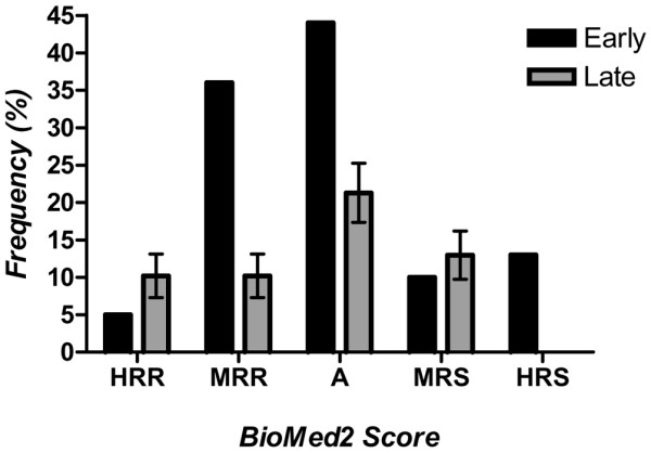
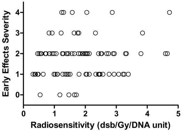
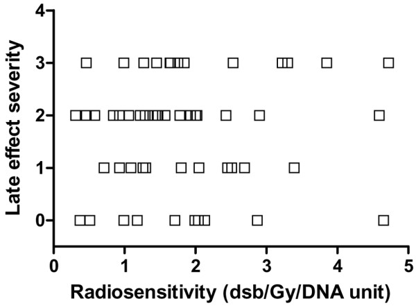
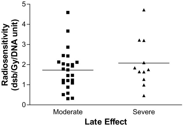
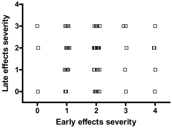
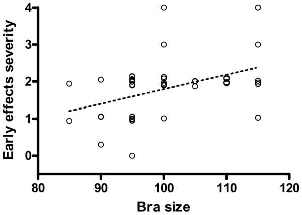
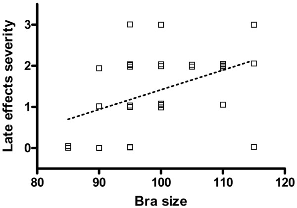
Results: Radiosensitivity values obtained using the in vitro test showed no relation with the acute or late adverse skin reactions observed. There was no evidence of a relation between acute and late normal tissue reactions assessed in the same patients. A positive relation was found between the treatment volume and both early and late side effects.

Conclusion: After radiation treatment, a number of cells containing major changes can have a long survival and disappear very slowly, becoming a chronic focus of immunological system stimulation. This stimulation can produce, in a stochastic manner, late radiation-related adverse effects of varying severity. Further research is warranted to identify the major determinants of normal tissue radiation response to make it possible to individualize treatments and improve the outcome of radiotherapy in cancer patients.


Figures

Figure 1
Figure 1
Frequency distribution of skin reactions in women with breast cancer treated postoperatively with radiotherapy. Reactions were classified as early (if observed at the end of the radiotherapy, 108 women) or late (if observed at the 7-year follow-up, 60 women). X-axis Radiation Therapy Oncology Group scoring system modified using the terminology proposed by Burnet (4): A, average; HRR, highly radioresistant; HRS, highly radiosensitive; MRR, moderately radioresistant; MRS, moderately radiosensitive.
Figure 2
Figure 2
Relation between severity of early radiotherapy-induced skin morbidity and lymphocyte molecular radiosensitivity. Skin morbidity in 108 women was assessed on the treated skin using the scoring system summarized in Table 1. Lymphocyte molecular radiosensitivity was measured as DNA double-strand breaks (dsb) by dose unit (Gy) and DNA unit (200 Mbp). bp, base pairs.
Figure 3
Figure 3
Relation between severity of late radiotherapy-induced skin morbidity and lymphocyte molecular radiosensitivity. Skin morbidity in 60 women was assessed on the treated skin using the scoring system summarized in Table 1. Lymphocyte molecular radiosensitivity was measured as DNA double-strand breaks (dsb) by dose unit (Gy) and DNA unit (200 Mbp). bp, base pairs.
Figure 4
Figure 4
Scatter-plot of quantified late skin reactions of patients and the corresponding in vitro radiosensitivity values. Horizontal solid lines are the mean values for each group of patients with breast cancer. Moderate (n = 38), women with reactions scored as highly or moderately radioresistant or as having an average response; severe (n = 12), patients with reactions scored as highly radiosensitive.
Figure 5
Figure 5
Relation between the severity of early and late side effects of radiotherapy for breast cancer. Women with breast cancer (n = 60) were treated postoperatively with radiotherapy and assessed using the scoring system summarized in Table 1.
Figure 6
Figure 6
Relation between estimated irradiation volumes and severity of early effects of radiotherapy for breast cancer. Irradiation volumes were estimated from the women's bra size, and the severity of the early effects were scored in the same women (n = 50). Dotted line shows the corresponding regression line (P < 0.001).
Figure 7
Figure 7
Relation between estimated irradiation volumes and severity of late effects of radiotherapy for breast cancer. Irradiation volumes were estimated from the women's bra size, and the late effects were scored in the same women (n = 33). Dotted line shows the corresponding regression line (P = 0.018).

References

    1. Johansson S, Svensson H, Denenkamp J. Dose response and latency for radiation induced fibrosis, edema and neuropathy in breast cancer patients. Int J Radiat Oncol Biol Phys. 2002;52:1207–1219. doi: 10.1016/S0360-3016(01)02743-2. - DOI - PubMed
    1. Van de Steene J, Vinh Hung V, Cutuli B, Storme G. Adjuvant radiotherapy for breast cancer: effects of longer follow-up. Radiother Oncol. 2004;72:35–43. doi: 10.1016/j.radonc.2004.04.004. - DOI - PubMed
    1. Van de Steene J, Soete G, Storme G. Adjuvant radiotherapy for breast cancer significantly improves overall survival: The missing link. Radiother Oncol. 2000;55:263–272. doi: 10.1016/S0167-8140(00)00204-8. - DOI - PubMed
    1. Burnet NG, Johansen J, Turesson I, Nyman J, Peacock JH. Describing patients' normal tissue reactions: concerning the possibility of individualising radiotherapy dose prescriptions based on potential predictive assays of normal tissue radiosensitivity. Steering Committee of the BioMed 2 European Union Concerted Action Programme on the Development of Predictive Tests of Normal Tissue Response to Radiation Therapy. Int J Cancer. 1998;79:606–613. doi: 10.1002/(SICI)1097-0215(19981218)79:6<606::AID-IJC9>3.0.CO;2-Y. - DOI - PubMed
    1. Brahme A. Individualizing cancer treatment: biological optimization models in treatment planning and delivery. Int J Radiat Oncol Biol Phys. 2001;49:327–337. doi: 10.1016/S0360-3016(00)01501-7. - DOI - PubMed

Publication types