. 2005;7(5):190-7.
doi: 10.1186/bcr1286. Epub 2005 Jul 12.

Myoepithelial cells: good fences make good neighbors

Affiliations

Myoepithelial cells: good fences make good neighbors

Melissa C Adriance et al. Breast Cancer Res. 2005.

Abstract

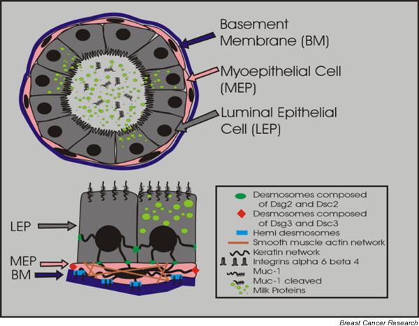
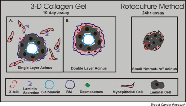
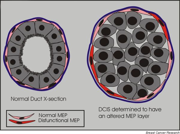
The mammary gland consists of an extensively branched ductal network contained within a distinctive basement membrane and encompassed by a stromal compartment. During lactation, production of milk depends on the action of the two epithelial cell types that make up the ductal network: luminal cells, which secrete the milk components into the ductal lumen; and myoepithelial cells, which contract to aid in the ejection of milk. There is increasing evidence that the myoepithelial cells also play a key role in the organizational development of the mammary gland, and that the loss and/or change of myoepithelial cell function is a key step in the development of breast cancer. In this review we briefly address the characteristics of breast myoepithelial cells from human breast and mouse mammary gland, how they function in normal mammary gland development, and their recently appreciated role in tumor suppression.


Figures

Figure 1
Figure 1
Cross-section of a bilayered duct. Secretory luminal cells (LEPs) are apically located to contractile myoepithelial cells (MEPs) and the basement membrane (BM). Milk proteins and cleaved Muc1 are secreted into the luminal space during lactation. Desmosomes containing desmoglein (Dsg)2 and desmocollin (Dsc)2 form between adjacent luminal cells and between adjacent LEPs and MEPs. Desmosomes between MEPs contain Dsg3 and Dsc3. MEPs as contractile cells contain smooth muscle actin and adhere to the BM via hemidesmosomes.
Figure 2
Figure 2
Three-dimensional culture method versus rotary culture. The methods shown utilize isolated purified human breast luminal and myoepithelial cells from reduction mammoplasty. In the three-dimensional culture method, coculture of purified luminal and myoepithelial cells in collagen I gel results in the formation of two different types of structures. The majority are (a) a single layer of cells that form acinar structures in which the secretion of laminin-1 by surrounding myoepithelial cells signals to luminal cells to polarize correctly, and the minority are (b) double layer acinar structures that are more reflective of the acinus in vivo. Gudjonsson and coworkers [15] showed that myoepithelial cells were able to induce correct luminal polarity via the synthesis of the basement membrane (BM) component laminin-1. In contrast, in the (c) rotary culture method, purified luminal and myoepithelial cells are grown in suspension. Acinar structures form, albeit at a smaller size compared with the three-dimensional method. Runswick and coworkers [5] showed that blocking desmosome adhesion via blocking peptides inhibited acinar formation.
Figure 3
Figure 3
DCIS myoepithelial cells exhibit an altered gene expression. In the normal breast myoepithelial cells (MEPs) are located between the luminal cells and the basement membrane. By their location they might act as a barrier to tumor invasion. In ductal carcinoma in situ (DCIS) the myoepithelial layer is still present; however, Allinen and coworkers [70] recently showed that there appears to be molecular differences between MEPs present in normal breast versus DCIS lesions.

References

    1. Ronnov-Jessen L, Petersen OW, Bissell MJ. Cellular changes involved in conversion of normal to malignant breast: importance of the stromal reaction. Physiol Rev. 1996;76:69–125. - PubMed
    1. Medina D. The mammary gland: a unique organ for the study of development and tumorigenesis. J Mammary Gland Biol Neoplasia. 1996;1:5–19. - PubMed
    1. Emerman JT, Vogl AW. Cell size and shape changes in the myoepithelium of the mammary gland during differentiation. Anat Rec. 1986;216:405–415. doi: 10.1002/ar.1092160310. - DOI - PubMed
    1. Radnor CJ. Myoepithelial cell differentiation in rat mammary glands. J Anat. 1972;111:381–398. - PMC - PubMed
    1. Runswick SK, O'Hare MJ, Jones L, Streuli CH, Garrod DR. Desmosomal adhesion regulates epithelial morphogenesis and cell positioning. Nat Cell Biol. 2001;3:823–830. doi: 10.1038/ncb0901-823. - DOI - PubMed

Publication types