. 2005 Oct;17(10):2723-37.
doi: 10.1105/tpc.105.034975. Epub 2005 Sep 16.

The RPN1 subunit of the 26S proteasome in Arabidopsis is essential for embryogenesis

Affiliations

The RPN1 subunit of the 26S proteasome in Arabidopsis is essential for embryogenesis

Vladimir Brukhin et al. Plant Cell. 2005 Oct.

Abstract

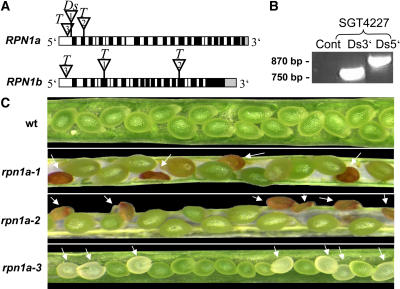
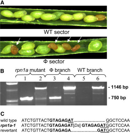
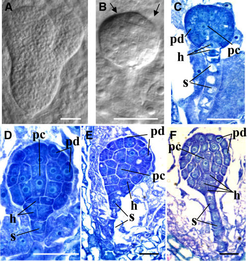
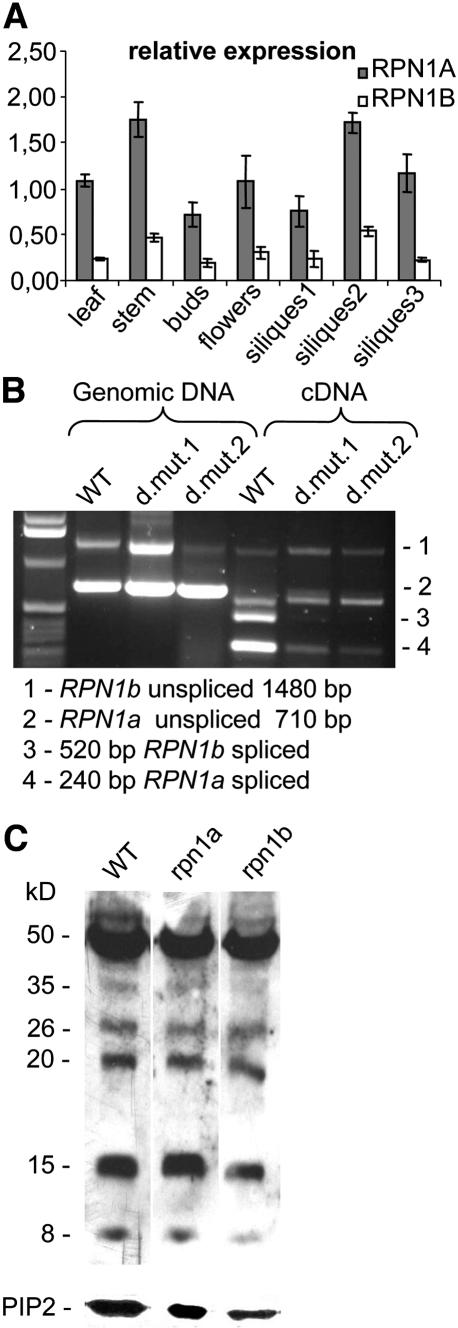
The 26S proteasome plays a central role in the degradation of regulatory proteins involved in a variety of developmental processes. It consists of two multisubunit protein complexes: the proteolytic core protease and the regulatory particle (RP). The function of most RP subunits is poorly understood. Here, we describe mutants in the Arabidopsis thaliana RPN1 subunit, which is encoded by two paralogous genes, RPN1a and RPN1b. Disruption of RPN1a caused embryo lethality, while RPN1b mutants showed no obvious abnormal phenotype. Embryos homozygous for rpn1a arrested at the globular stage with defects in the formation of the embryonic root, the protoderm, and procambium. Cyclin B1 protein was not degraded in these embryos, consistent with cell division defects. Double mutant plants (rpn1a/RPN1a rpn1b/rpn1b) produced embryos with a phenotype indistinguishable from that of the rpn1a single mutant. Thus, despite their largely overlapping expression patterns in flowers and developing seeds, the two isoforms do not share redundant functions during gametogenesis and embryogenesis. However, complementation of the rpn1a mutation with the coding region of RPN1b expressed under the control of the RPN1a promoter indicates that the two RPN1 isoforms are functionally equivalent. Overall, our data indicate that RPN1 activity is essential during embryogenesis, where it might participate in the destruction of a specific set of protein substrates.


Figures

Figure 1.
Figure 1.
RPN1 Mutant Alleles and rpn1a Phenotypes. (A) Schematic representation of the RPN1a and RPN1b genes. RPN1a: insertion of the Ds element (triangle 1) in SGT4227 (rpn1a-1) into the ATG start codon in the first RPN1a exon; the T-DNA insertion (triangle 2) in SALK_12604 (rpn1a-2) disrupts the second exon of RPN1a; the T-DNA insertion in SALK_30503 (rpn1a-3) lies 300 bp upstream of the RPN1a gene coding region. RPN1b: the T-DNA insertion in SALK_115981 (rpn1b-1) disrupts the 5th exon and in SALK_061211 (rpn1b-2) the 17th exon of RPN1b, respectively; in SALK_023490 (rpn1b-3), the T-DNA inserted into the promoter region. Black boxes indicate exons, white boxes introns, and gray boxes the 3′ untranslated region. (B) PCR analysis of the sequences spanning the 5′ and 3′ junctions of the Ds insertion in rpn1a-1. (C) rpn1a mutants are embryo lethal. Wild-type siliques show full seed set, while in rpn1a-1, rpn1a-2, and rpn1a-3 heterozygous plants, one-quarter of the seeds abort (arrows).
Figure 2.
Figure 2.
Analysis of rpn1a-1 Revertant Sectors. (A) Phenotype of a mosaic hybrid plant obtained from the cross between a Ac4/Ac4 female and a rpn1a-1/RPN1a male, showing siliques from a wild-type (top panel) and a phenotypic sector with aborted seeds (arrows, bottom panel). (B) PCR analysis of genomic DNA from the phenotypic sector (lanes 3 and 4), the wild-type sector (lanes 5 and 6), and the rpn1a-1/RPN1a control plant (lanes 1 and 2). A fragment of 750 bp specific to the Ds insertion in the rpn1a-1 allele is missing due to excision of the Ds element (lane 5); the 1146-bp fragment specific to the Ds element is present in all samples shown. (C) DNA sequences flanking the rpn1a-1 Ds insertion, which created a typical 8-bp direct target site duplication. The ATG start codon is underlined. In the revertant allele, one nucleotide was deleted at the 5′ junction and five nucleotides at the 3′ junction, leaving a typical Ds footprint of 2 bp. Excision removed the large insertion restoring the function of RPN1a.
Figure 3.
Figure 3.
Seed and Embryo Development in rpn1a-1/RPN1a Plants. (A) and (B) Cleared seeds from the same silique: torpedo stage of a wild-type seed (A), aborted seed with the embryo arrested at the globular stage (B). Note the irregular protoderm layer (arrows). (C) to (F) Toluidine blue–stained sections of a wild-type embryo at the early globular stage (C) and rpn1a-1 mutant embryos at the globular stage with abnormalities described in the text ([D] to [F]). h, derivatives of hypophyseal cell; pc, procambial cells; pd, protoderm; s, suspensor. Bars = 25 μm in (A) and 50 μm in (B) to (F).
Figure 4.
Figure 4.
Expression and Function of the RPN1 Genes in the Wild Type and Mutants. (A) Relative RPN1a and RPN1b transcript accumulation in wild-type tissues. Both transcripts were detected in vegetative and reproductive tissues. RPN1a was consistently expressed at higher levels than RPN1b. Data are presented as values relative to 18S RNA expression. Identical results were obtained using ACTIN1 as a reference gene (data not shown). Error bars represent sd on the basis of three biological replicates. (B) RT-PCR analysis for expression of the RPN1a and RPN1b genes in double mutants heterozygous for rpn1a-1 and homozygous for rpn1b-2 and rpn1b-3, respectively (here indicated as d.mut.1 and d.mut.2). No RPN1b mRNA is detected in the double mutants, and RPN1a mRNA levels are reduced compared with the wild type, likely due to heterozygosity. (C) Polyubiquinated proteins in siliques from the wild type, rpn1a/RPN1a, and rpn1b/rpn1b mutants. Total protein was separated on a gel and transferred to a membrane, and polyubiquinated proteins were detected using a polyclonal antiubiquitin antibody. The bottom panel shows the AtPIP2b plasma membrane protein as loading control. The position of molecular mass marker is shown at the left (in kD).
Figure 5.
Figure 5.
In Situ Detection of RPN1a mRNA in Developing Flowers, Seeds, and Siliques and cyclinB1;1 Expression in the rpn1a-1 Mutant. (A) to (D) Longitudinal sections of a floral meristem and flower buds at various stages. (E) and (F) Embryo at the octant stage. (G) and (H) Embryo at the globular stage. (I) and (J) Embryo at the late heart stage. (K) and (L) Embryo at the walking stick stage. (M) to (O) Fragment of a silique. (P) Tetrads of microspores. The hybridization signal appears as dark staining. (Q) Embryo carrying at least one wild-type RPN1a allele. The cyclinB1;1-GUS fusion protein is degraded and barely detectable in the embryo (arrow). (R) rpn1a-1 homozygous mutant embryo from the same silique as in (Q) accumulates cyclinB1;1-GUS fusion protein (arrow). AS, antisense probe; S, sense probe; ch, chalaza; ov, ovules. Bars = 25 μm in (A), (B), (E) to (H), (O), and (P) and 50 μm in (C), (D), and (I) to (N).
Figure 6.
Figure 6.
In Situ Detection of RPN1b mRNA in Developing Flowers, Seeds, and Siliques. (A) and (B) Longitudinal sections of a flower. (C) and (D) Embryo at the octant stage. (E) and (F) Embryo at the globular stage. (G) and (H) Embryo at the torpedo stage. (I) and (J) Fragment of a silique. Hybridization signal appears as dark staining. AS, antisense probe; S, sense probe. Bars = 25 μm in (C) to (F) and 50 μm in (A), (B), and (G) to (J).

References

    1. Alonso, J.M., et al. (2003). Genome-wide insertional mutagenesis of Arabidopsis thaliana. Science 301, 653–657. - PubMed
    1. Altschul, S.F., Madden, T.L., Schaffer, A.A., Zhang, J., Zhang, Z., Miller, W., and Lipman, D.J. (1997). Gapped BLASTand PSI-BLAST: A new generation of protein database search programs. Nucleic Acids Res. 25, 3389–3402. - PMC - PubMed
    1. Arabidopsis Genome Initiative (2000). Analysis of the genome sequence of the flowering plant Arabidopsis thaliana. Nature 408, 796–815. - PubMed
    1. Bailly, E., and Reed, S.U. (1999). Functional characterization of Rpn3 uncovers a distinct 19S proteasomal subunit requirement for ubiquitin-dependent proteolysis of cell cycle regulatory proteins in budding yeast. Mol. Cell. Biol. 19, 6872–6890. - PMC - PubMed
    1. Blanc, G., Barakat, A., Guyot, R., Cooke, R., and Delseny, M. (2000). Extensive duplication and reshuffling in the Arabidopsis genome. Plant Cell 12, 1093–1101. - PMC - PubMed

Publication types

MeSH terms