. 2005 Nov 8;102(45):16128-32.
doi: 10.1073/pnas.0506586102. Epub 2005 Sep 16.

Direct observation of thymine dimer repair in DNA by photolyase

Affiliations

Direct observation of thymine dimer repair in DNA by photolyase

Ya-Ting Kao et al. Proc Natl Acad Sci U S A. .

Abstract

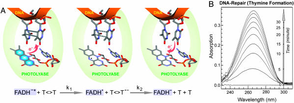
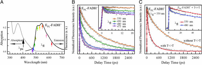
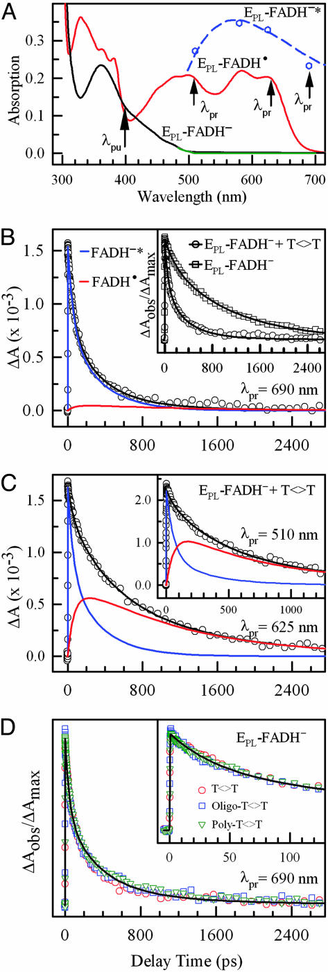
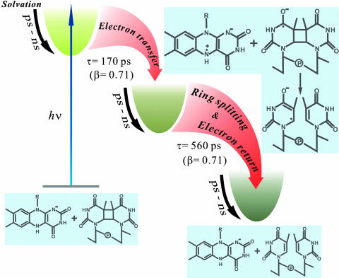
Photolyase uses light energy to split UV-induced cyclobutane dimers in damaged DNA, but its molecular mechanism has never been directly revealed. Here, we report the direct mapping of catalytic processes through femtosecond synchronization of the enzymatic dynamics with the repair function. We observed direct electron transfer from the excited flavin cofactor to the dimer in 170 ps and back electron transfer from the repaired thymines in 560 ps. Both reactions are strongly modulated by active-site solvation to achieve maximum repair efficiency. These results show that the photocycle of DNA repair by photolyase is through a radical mechanism and completed on subnanosecond time scale at the dynamic active site, with no net change in the redox state of the flavin cofactor.


Figures

Fig. 1.
Fig. 1.
Repair of damaged DNA by photolyase. (A) Schematic representation of DNA-repair processes by photolyase through an electron-transfer radical mechanism. The key catalytic reactions, charge separation (k1) and ring splitting (k2), are given at the bottom. Our study follows the evolution of the flavin cofactor. (B) The steady-state repair of dinucleotide thymine dimer (200 μM) by photolyase (10 μM) by illumination of the reaction mixture under 360-nm light. The thymine monomer formation was detected by increase in absorption at 260 nm.
Fig. 2.
Fig. 2.
Ultrafast fluorescence spectroscopy of a photolyase in the absence and presence of substrate. (A) Absorption and emission spectra of photolyase containing FADH cofactor (EPL-FADH) and no second chromophore in the absence of substrate. The pump wavelength was fixed at 400 nm for all experiments. Nine fluorescence wavelengths were gated from the blue side to the red side. (B) Four typical gated fluorescence transients, with systematic decays from 475 to 550 nm, reflecting significant solvation at the active site. Inset shows fluorescent transients at early time points. (C) The fluorescence transients at 550 nm with and without the substrate thymine dimer. The enzyme concentration was 0.4 mM, and the substrate concentration was 8 mM. Inset shows the fluorescence transients at 480 and 550 nm in the presence of substrate. The initial ultrafast solvation at 480 nm is still present.
Fig. 3.
Fig. 3.
Determination of forward and back electron transfer in photolyase photocycle by ultrafast absorption spectroscopy. (A) Absorption spectra of EPL-FADH· (red) and EPL-FADH* (blue), as well as EPL-FADH before (black) and after (green) the repair experiment. The absorption profile of EPL-FADH* was obtained from ref. and calibrated by our four transient-absorption data at 510, 580, 620, and 690 nm, relative to FADH·. (B) The absorption transient probed at 690 nm, showing a dominant contribution of FADH* decay (95%) with a minor signal from FADH·. The enzyme concentration is 0.4 mM, and the substrate concentration is 8 mM. Inset shows the drastically different dynamics with and without the substrate. (C) Absorption transients probed at 625 and 510 nm (Inset) show both FADH* and the intermediate FADH· dynamics. (D) Absorption transients probed at 690 nm for a series of substrates, dinucleotide thymine dimer, and oligo(dT)12–18 and poly(dT) with dimer. The oligomer concentration is 1 mM, and the total dimer concentration in poly(dT) is ≈5 mM. Inset shows the early time points of the reaction.
Fig. 4.
Fig. 4.
Evolution of catalytic reactions of DNA repair by photolyase along the coordinate. Active-site solvation strongly modulates the charge-separation, ring-splitting, and electron-return processes, resulting in slow charge separation (170 ps) and a stretched-single-exponential-decay dynamics (β = 0.71). The charge recombination must be slower than the complete ring splitting (560 ps) to eliminate possible ring reclosure and achieve a maximum-repair quantum yield (0.87).

References

    1. Brash, D. E. (1997) Trends Genet. 13, 410–414. - PubMed
    1. Rupert, C. S., Goodgal, S. H. & Herriott, R. M. (1958) J. Gen. Physiol. 41, 451–471. - PMC - PubMed
    1. Sancar, A. (2003) Chem. Rev. 103, 2203–2237. - PubMed
    1. Park, H. W., Kim, S. T., Sancar, A. & Deisenhofer, J. (1995) Science 268, 1866–1872. - PubMed
    1. Sancar, G. B., Jorns, M. S., Payne, G., Fluke, D. J., Rupert, C. S. & Sancar, A. (1987) J. Biol. Chem. 262, 492–498. - PubMed

Publication types