. 2005 Oct;139(2):701-12.
doi: 10.1104/pp.105.065607. Epub 2005 Sep 16.

Cell-to-cell movement of green fluorescent protein reveals post-phloem transport in the outer integument and identifies symplastic domains in Arabidopsis seeds and embryos

Affiliations

Cell-to-cell movement of green fluorescent protein reveals post-phloem transport in the outer integument and identifies symplastic domains in Arabidopsis seeds and embryos

Ruth Stadler et al. Plant Physiol. 2005 Oct.

Abstract

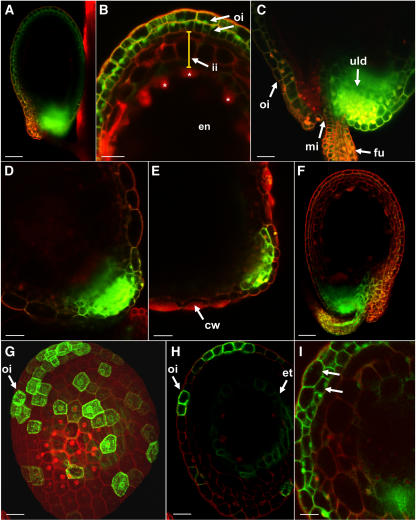
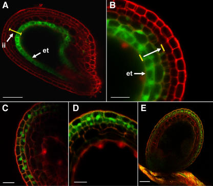
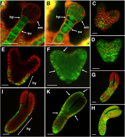
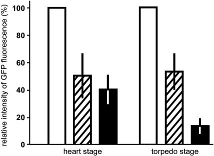
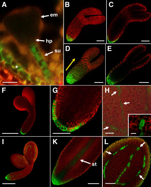
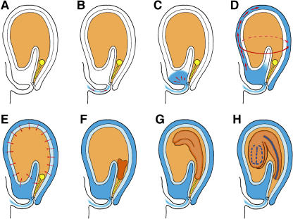
Developing Arabidopsis (Arabidopsis thaliana) seeds and embryos represent a complex set of cell layers and tissues that mediate the transport and partitioning of carbohydrates, amino acids, hormones, and signaling molecules from the terminal end of the funicular phloem to and between these seed tissues and eventually to the growing embryo. This article provides a detailed analysis of the symplastic domains and the cell-to-cell connectivity from the end of the funiculus to the embryo, and within the embryo during its maturation. The cell-to-cell movement of the green fluorescent protein or of mobile and nonmobile green fluorescent protein fusions was monitored in seeds and embryos of plants expressing the corresponding cDNAs under the control of various promoters (SUC2, SUC3, TT12, and GL2) shown to be active in defined seed or embryo cell layers (SUC3, TT12, and GL2) or only outside the developing Arabidopsis seed (AtSUC2). Cell-to-cell movement was also analyzed with the low-molecular-weight fluorescent dye 8-hydroxypyrene-1,3,6-trisulfonate. The analyses presented identify a phloem-unloading domain at the end of the funicular phloem, characterize the entire outer integument as a symplastic extension of the phloem, and describe the inner integument and the globular stage embryo plus the suspensor as symplastic domains. The results also show that, at the time of hypophysis specification, the symplastic connectivity between suspensor and embryo is reduced or interrupted and that the embryo develops from a single symplast (globular and heart stage) to a mature embryo with new symplastic domains.


Figures

Figure 1.
Figure 1.
Analysis of the symplastic connectivity and of post-phloem GFP movement in the outer integument of Arabidopsis seeds. A to E, Seeds from AtSUC2 promoter/GFP plants. A, Overview of the GFP fluorescence in a seed from the fifth silique (2 d old; globular stage of the embryo; bar = 50 μm). B, Magnification of the tip region of a seed similar to that shown in A. GFP fluorescence is detected in both cell layers of the outer integument (oi). No GFP fluorescence is seen in any of the three cell layers of the inner integument (ii; yellow bar) or in the endosperm (en). Some of the peripheral endosperm nuclei are marked with asterisks (bar = 25 μm). C, Magnification of the basal unloading (uld) domain at the end of the funiculus (fu) of a seed similar to that shown in A. Weak GFP fluorescence is seen in both cell layers of the outer integument (oi; mi, micropyle; bar = 25 μm). D, GFP fluorescence in a seed from the twelfth silique (6 d old; upturned-U stage of the embryo). At this stage, GFP is still unloaded from the funicular vascular bundle, but post-phloem movement of GFP in the outer integument is no longer detected (bar = 25 μm). E, GFP fluorescence in a seed from the fifteenth silique (10 d old; mature embryo). GFP fluorescence is seen only in few cells of the unloading domain. At this stage, cell wall (cw) thickening and development of the testa have already started (bar = 25 μm). F, Seed from an AtSUC2 promoter/GFP-sporamin plant. GFP-sporamin is unloaded from the vascular bundle at the end of the funiculus, but post-phloem movement in the outer integument is not detected (2 d old; globular stage of the embryo; bar = 40 μm). G and H, Seed from an AtGL2 promoter/tmGFP9 plant. G, View of the seed from the outside showing AtGL2 promoter activity in individual cells of the outer integument (oi; bar = 20 μm). H, Optical section through the seed shown in G confirming the AtGL2 promoter activity in the outermost cell layer of the outer integument (oi). A weak activity of the AtGL2 promoter is also seen in the innermost layer of the inner integument, the endothelium (et; bar = 20 μm). I, Seed of an AtGL2 promoter/GFP plant. GFP fluorescence is seen in both layers of the outer integument (arrows) showing that GFP can move cell to cell not only within the outer cell layer but also from the outer into the inner cell layer (bar = 25 μm). All images represent CLSM images. G is a maximum projection of a picture stack. All others represent optical sections. Red color results from propidium iodide staining of cell walls.
Figure 2.
Figure 2.
Analysis of the symplastic connectivity between the inner and outer integuments of Arabidopsis seeds. A and B, Analysis with AtTT12 promoter/tmGFP9 plants. C to E, Analysis with HPTS. A, Seed of an AtTT12 promoter/tmGFP9 plant. The fluorescence of the membrane-bound GFP is restricted to the endothelium (et). No tmGFP9 fluorescence is detected in the two other cell layers of the inner integument (ii; yellow bar; bar = 50 μm). B, Seed of an AtTT12 promoter/GFP plant. Strong GFP fluorescence is seen in the endothelial (et) layer, where the AtTT12 promoter is active (see A), but clearly GFP can diffuse into the two adjacent cell layers (direction of the white arrow). The two yellow bars show the inner and outer borders of the inner integument (scale bar = 20 μm). Both images represent optical sections (CLSM). Red color results from propidium iodide staining of cell walls. C, Optical section through an Arabidopsis wild-type seed 120 min after HPTS loading (bar = 20 μm). D, Optical section through an Arabidopsis wild-type seed 4 h after HPTS loading (bar = 20 μm). E, Optical section through an Arabidopsis wild-type seed 5 h after HPTS loading (bar = 50 μm). All images represent CLSM images. All images represent optical sections. Red color results from propidium iodide staining of cell walls.
Figure 3.
Figure 3.
Symplastic connectivity in the suspensor and the developing Arabidopsis embryo. A, Globular stage embryo (em) and suspensor (su) of an AtSUC3 promoter/tmGFP9 plant. Green tmGFP9-derived fluorescence is detected in all cells of the suspensor and in the hypophysis (hp). The asterisk marks the endothelium, where the AtSUC3 promoter is also active (bar = 25 μm). B, Globular stage embryo (em), hypophysis (hp), and suspensor (su) of an AtSUC3 promoter/GFP plant. In contrast to A, green GFP-derived fluorescence is detected also in all cells of the embryo. The asterisk marks the endothelium, where the AtSUC3 promoter is also active (bar = 25 μm). C, View of an intact early heart-stage embryo of an AtGL2 promoter/tmGFP9 plant showing tmGFP9 fluorescence only in distinct cells in the hypocotyl region (bar = 20 μm). D, View of an intact early heart-stage embryo of an AtGL2 promoter/GFP plant showing GFP in all cells. Bright green dots show GFP accumulation in nuclei (bar = 20 μm). E, Optical section of the embryo shown in C showing that tmGFP9 fluorescence is restricted to the hypocotyl (hy) epidermis (bar = 20 μm). F, Optical section of the embryo shown in D showing GFP in all epidermis cells (arrows), but also in all underlying cell layers (bar = 20 μm). G, View of a midtorpedo-stage embryo of an AtGL2 promoter/tmGFP9 plant. tmGFP9 fluorescence is seen only in rows of cells in the hypocotyl and the lower part of the cotyledons (bar = 40 μm). H, View of an intact midtorpedo-stage embryo of an AtGL2 promoter/GFP plant. GFP fluorescence is seen in all cells (bar = 40 μm). I, Optical section of the embryo shown in G showing that tmGFP9 fluorescence is restricted to the hypocotyl (hy) epidermis (bar = 40 μm). K, Optical section of the embryo similar to that shown in H showing strong GFP fluorescence in all cells of the epidermis (arrows) and in the root tip. GFP can still move into the underlying cell layers of the embryo, but movement into these cells is clearly reduced compared to the heart-stage embryo shown in F (bar = 40 μm). All images represent CLSM pictures. C, D, G, and H are maximum projections of picture stacks; all others represent optical sections. Red color shows propidium iodide-stained cell walls in A and B and chlorophyll autofluorescence in all other images.
Figure 4.
Figure 4.
Quantification of the intensity of GFP fluorescence in different areas of heart-stage and torpedo-stage embryos of AtGL2 promoter/GFP plants. The GFP fluorescence (497–526 nm; mean ± sd) from four to six images was determined in three different areas by integration. The selected areas were (1) epidermis from the hypocotyl (white bars), where the AtGL2 promoter is known to be active (see Fig. 3, E and I); (2) epidermis from the emerging cotyledons (hatched bars), where the AtGL2 promoter was shown not to be active (see Fig. 3, E and I); and (3) ground tissue cells from the emerging cotyledons (black bars). All values were normalized to the same area and the intensity at the site of AtGL2 promoter activity was always set to 100%. The hatched bars show that the intensity of GFP movement within the epidermis is the same in heart-stage and in torpedo-stage embryos. The black bars demonstrate that movement of GFP from the epidermis into the underlying ground tissue is reduced by a factor of 3 during the transition from heart stage to torpedo stage.
Figure 5.
Figure 5.
Initiation and formation of symplastic units during embryo maturation. A, Optical section through a triangular stage embryo (em) of an AtSUC3 promoter/GFP plant. GFP fluorescence is seen in the endothelial layer of the inner integument (asterisk) and in the suspensor (su). Movement of GFP from the suspensor into the embryo or the hypophysis (hp) is no longer detected at this stage (bar = 25 μm). B and C, Confocal projection (B) or optical section (C) of an AtSUC3 promoter/tmGFP9 embryo in the midtorpedo stage. B, View of the intact embryo. tmGFP9 fluorescence is seen only in the root tip (bar = 50 μm). C, tmGFP9 fluorescence is restricted to the root-tip epidermis (bar = 50 μm). D and E, Confocal projection (D) or optical section (E) of an AtSUC3 promoter/GFP embryo in the midtorpedo stage. D, View of the intact embryo shows a gradient of GFP diffusing from the root tip toward the apex (yellow arrow; bar = 50 μm). E, GFP does move from the root-tip epidermis into the adjacent cell layers of the embryo, but, as in Figures 3K and 5D, diffusion seems to be better within the epidermis (bar = 50 μm). F to H, View from the outside (confocal projection) of the almost mature embryo of an AtSUC3 promoter/tmGFP9 plant (F) or optical sections from the embryo root tip (G) or the cotyledon (H). F, tmGFP9 fluorescence is seen only in the root tip. The fluorescence shown in H is weaker than that in the root tip and is not seen in this image (bar = 200 μm). G, tmGFP9 fluorescence is confined to the epidermis of the root tip (bar = 50 μm). H, tmGFP9 fluorescence is seen in individual cells of the cotyledon (arrows). Due to the low expression of tmGFP9 in these cells, the cell borders are difficult to visualize. The inset shows a single cell at slightly higher magnification (bar = 25 μm; inset, 10 μm). I to L, View from the outside (confocal projection) of the almost mature embryo of an AtSUC3 promoter/GFP plant (I) or optical sections from the embryo root tip (K) or the cotyledon (L). I, GFP fluorescence is seen in distinct cells of the cotyledons and in the root tip (bar = 200 μm). K, GFP can move from the cells of the root-tip epidermis into all adjacent cells, but movement of GFP is seen primarily in the developing stele (st) in the center of the hypocotyl (bar = 50 μm). L, GFP fluorescence is seen in individual cells on the cotyledon (arrows). No movement of GFP out of these cells can be detected (bar = 50 μm). Red color shows chlorophyll autofluorescence in all images.
Figure 6.
Figure 6.
Model showing the symplastic domains in a developing Arabidopsis seed and the deduced path of assimilates. A, All maternal tissues of the seed are shown in white, the endosperm in light orange, and the suspensor and the embryo in yellow. B, Flux of assimilates in the vascular bundle of the funiculus. This bundle ends in the testa proximal to the micropyle. C, Unloading into the post-phloem unloading domain. D, Movement within the outer integument (red arrows), which forms a symplastic extension of the vascular tissue. E, Centripetal transport (red arrows) from the outer integument toward the endosperm over two apoplastic borders. In the young seed, the suspensor and the embryo form a single symplastic domain (Fig. 3, A and B). F, During the early heart stage. the symplastic continuity between embryo and suspensor is interrupted (Fig. 5A) and the embryo forms a single symplastic domain. G, During the transition from heart to midtorpedo stage, the symplastic connectivity between epidermis and inner cell layers of the embryo is gradually reduced. The symplastic connectivity between the cells of the epidermis is high. H, In mature embryos, the developing stele in the root and the hypocotyl begins to form an independent domain. Cells within the stele show high symplastic connectivity, allowing the movement of GFP synthesized in the root-tip epidermis (Fig. 5, F and G). The symplastic connectivity between developing stele and root cortex is low and significant movement of GFP out of the stele is not observed. At the same time, individual cells along the leaf vascular bundles show reduced symplastic connectivity to their adjacent cells. GFP synthesized in these cells (Fig. 5H) does not move out of these cells (Fig. 5L).

References

    1. Becker D, Kemper E, Schell J, Mastersen R (1992) New plant binary vectors with selectable markers located proximal to the left T-DNA border. Plant Mol Biol 20: 1195–1197 - PubMed
    1. Berleth T, Jürgens G (1993) The role of the MONOPTEROS gene in organizing the basal body region of the Arabidopsis embryo. Development 118: 575–587
    1. Blilou I, Xu J, Wildwater M, Willemsen V, Paponov I, Friml J, Heidstra R, Aida M, Palme K, Scheres B (2005) The PIN auxin efflux facilitator network controls growth and patterning in Arabidopsis roots. Nature 433: 39–44 - PubMed
    1. Bowman JL (1993) Embryogenesis. In JL Bowman, ed, Arabidopsis: An Atlas of Morphology and Development. Springer-Verlag, New York, pp 351–401
    1. Debeaujon I, Peeters AJM, Léon-Kloosterziel KM, Koornneef M (2001) The TRANSPARENT TESTA12 gene of Arabidopsis encodes a multidrug secondary transporter-like protein required for flavonoid sequestration in vacuoles of the seed coat endothelium. Plant Cell 13: 853–872 - PMC - PubMed

Publication types

MeSH terms