. 2005 Dec;89(6):4201-10.
doi: 10.1529/biophysj.105.068726. Epub 2005 Sep 16.

Amyloid fibril formation can proceed from different conformations of a partially unfolded protein

Affiliations

Amyloid fibril formation can proceed from different conformations of a partially unfolded protein

Martino Calamai et al. Biophys J. 2005 Dec.

Abstract

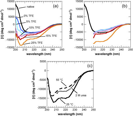
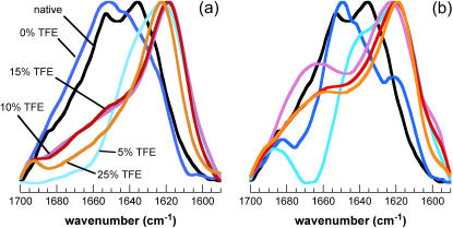
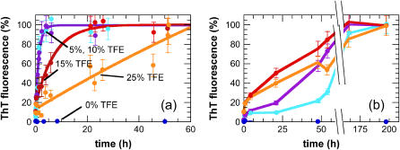
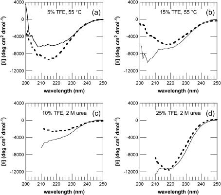
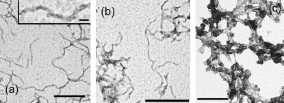
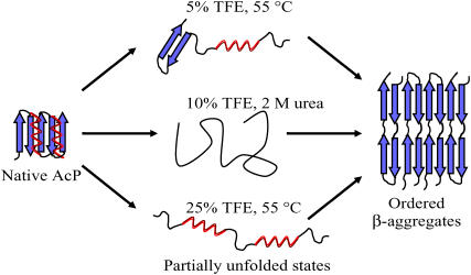
Protein misfolding and aggregation are interconnected processes involved in a wide variety of nonneuropathic, systemic, and neurodegenerative diseases. More generally, if mutations in sequence or changes in environmental conditions lead to partial unfolding of the native state of a protein, it will often aggregate, sometimes into well-defined fibrillar structures. A great deal of interest has been directed at discovering the characteristic features of metastable partially unfolded states that precede the aggregated states of proteins. In this work, human muscle acylphosphatase (AcP) has been first destabilized, by addition of urea or by means of elevated temperatures, and then incubated in the presence of different concentrations of 2,2,2, trifluoroethanol ranging from 5% to 25% (v/v). The results show that AcP is able to form both fibrillar and nonfibrillar aggregates with a high beta-sheet content from partially unfolded states with very different structural features. Moreover, the presence of alpha-helical structure in such a state does not appear to be a fundamental determinant of the ability to aggregate. The lack of ready aggregation under some of the conditions examined here is attributable primarily to the intrinsic properties of the solutions rather than to specific structural features of the partially unfolded states that precede aggregation. Aggregation appears to be favored when the solution conditions promote stable intermolecular interactions, particularly hydrogen bonds. In addition, the structures of the resulting aggregates are largely independent of the conformational properties of their soluble precursors.


Figures

FIGURE 1
FIGURE 1
Far-UV CD spectra of different initial states from which AcP aggregates. Spectra were acquired 1 min after the addition of different concentrations of TFE to AcP samples preincubated at 55°C (a) and in 2 M urea at 25°C (b). In b, the F94L mutant was used instead of wild-type (wt) AcP (see text). Black indicates the native state at 25°C in the absence of TFE and urea; the colors indicate different TFE concentrations added to the destabilized state at 55°C or 2 M urea: blue, 0% TFE; cyan, 5% TFE; purple, 10% TFE; red, 15% TFE; orange, 25% TFE. (c) Comparison of the spectra of samples at 25°C (solid line), 55°C (dashed line), and in 2 M urea, 25°C (dotted line), all obtained in the presence of 25% TFE.
FIGURE 2
FIGURE 2
ATR-FTIR spectra of different aggregated states of AcP. Amide I′ spectra were acquired 1 week after the addition of different concentrations of TFE to samples preincubated at 55°C (a) and in 2 M urea at 25°C (b). In b, the F94L mutant was used instead of wt AcP. The color code is the same as in Fig. 1. Spectra were normalized to the minimum and maximum absorbance values in each experiment.
FIGURE 3
FIGURE 3
Aggregation kinetics of AcP from different partially unfolded states. Aggregation was monitored by ThT fluorescence after the addition of different concentrations of TFE to samples preincubated at 55°C (a) and in 2 M urea, 25°C (b). In b, the F94L mutant was used instead of wt AcP. The color code is the same as in Fig. 1. The data points were normalized to attribute 100% intensity to the maximum value of ThT fluorescence intensity observed at the end of each experiment. The solid lines in a represent the single exponential functions which provide the best fits to the data.
FIGURE 4
FIGURE 4
Far-UV CD spectra of AcP before and after aggregation. Spectra were acquired after 1 min (solid line), 1 day (dotted line, a and b), or 1 week (dotted line, c and d) after the addition of different concentrations of TFE to samples preincubated at 55°C (a and b) and in 2 M urea (c and d). In c and d, the F94L mutant was used instead of wt AcP. The samples were incubated in 5% (a), 10% (c), 15% (b), and 25% (d) TFE. A similar shift of the peak to wavelengths between 210 and 220 nm was observed also for the samples incubated in 15–25% TFE at 55°C, and 5–10% TFE in 2 M urea.
FIGURE 5
FIGURE 5
Aggregates of AcP observed by TEM. The protein was incubated in 5% (a) and 15% TFE (b) at 55°C or in 10% TFE and 2 M urea at 25°C (c), and images were acquired after 1 week. In c, the F94L mutant was used instead of wt AcP. An expanded image is shown in the inset (a). The scale bars at the bottom of a and c correspond to 100 nm; those in b and in the inset correspond to 50 nm and 10 nm, respectively.
FIGURE 6
FIGURE 6
Schematic representation of the aggregation process of AcP from different partially unfolded states. The globular native state of AcP is converted into a partially unfolded state that can contain a mixture of α-helical and β-sheet structure (5% TFE, 55°C), a high content of α-helical (25% TFE, 55°C) or disordered (F94L, 10% TFE, 2 M urea, 25°C) structure. Despite the differences in the conditions, all of these partially unfolded states can be converted into ordered fibrillar aggregates containing a highly organized β-sheet structure.

References

    1. Dobson, C. M. 2003. Protein folding and misfolding. Nature. 426:884–890. - PubMed
    1. Walsh, D. M., and D. J. Selkoe. 2004. Oligomers on the brain: the emerging role of soluble protein aggregates in neurodegeneration. Protein Pept. Lett. 11:213–228. - PubMed
    1. Buxbaum, J. N. 2004. The systemic amyloidoses. Curr. Opin. Rheumatol. 16:67–75. - PubMed
    1. Pepys, M. B. 2001. Pathogenesis, diagnosis and treatment of systemic amyloidosis. Philos. Trans. R. Soc. Lond. B Biol. Sci. 356:203–210 (discussion 210–201). - PMC - PubMed
    1. Stefani, M., and C. M. Dobson. 2003. Protein aggregation and aggregate toxicity: new insights into protein folding, misfolding diseases and biological evolution. J. Mol. Med. 81:678–699. - PubMed

Publication types

LinkOut - more resources