

Whole body PET/CT for initial staging of choroidal melanoma
- PMID: 16170114
- PMCID: PMC1772897
- DOI: 10.1136/bjo.2005.069823
Whole body PET/CT for initial staging of choroidal melanoma
Abstract
Aim: To investigate the value of whole body positron emission tomography/computed tomography (PET/CT) in screening for metastatic choroidal melanoma in patients initially diagnosed with choroidal melanoma.
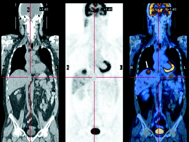
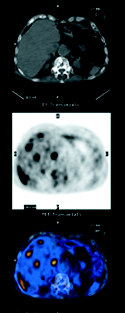
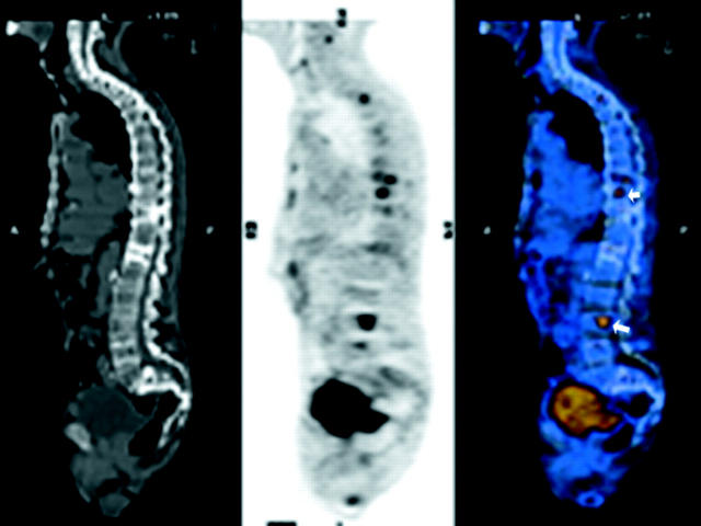
Methods: 52 patients with choroidal melanoma underwent whole body PET/CT as part of their metastatic investigation. PET/CT scans were used as a screening tool at the time of their initial diagnosis. A physical examination, liver function tests, and a baseline chest x ray were also obtained. PET/CT images (utilising intravenous18-fluoro-2-deoxyglucose (FDG)) were studied for the presence of metastatic melanoma. The standards for reference were further imaging and/or subsequent biopsies.
Results: Two of 52 (3.8%) patients were found to have metastatic melanoma before treatment. The most common sites for metastases were the liver (100%), bone (50%), and lymph nodes (50%). Brain involvement was also present in one patient. One patient (50%) had involvement of multiple sites. Haematological liver enzyme assays were normal in both patients. PET/CT showed false positive results in three patients (5.7%) when further evaluated by histopathology and/or additional imaging. In seven patients (13.4%) PET/CT imaging detected benign lesions in the bone, lung, lymph nodes, colon, and rectum.
Conclusion: PET/CT imaging can be used as a screening tool for the detection and localisation of metastatic choroidal melanoma. Liver enzyme assays did not identify liver metastases, while PET/CT revealed both hepatic and extrahepatic metastatic melanoma. PET/CT imaging may improve upon the conventional methods of screening for detection of metastatic disease in patients initially diagnosed with choroidal melanoma.
Figures
Similar articles
-
Whole body positron emission tomography/computed tomography staging of metastatic choroidal melanoma.Am J Ophthalmol. 2005 Aug;140(2):193-9. doi: 10.1016/j.ajo.2005.02.051. Am J Ophthalmol. 2005. PMID: 15992753 Clinical Trial.
-
Initial PET/CT staging for choroidal melanoma: AJCC correlation and second nonocular primaries in 333 patients.Eur J Ophthalmol. 2012 Mar-Apr;22(2):236-43. doi: 10.5301/ejo.5000049. Eur J Ophthalmol. 2012. PMID: 21959680
-
Whole-body 18 FDG PET/CT imaging for lymph node and metastatic staging of conjunctival melanoma.Br J Ophthalmol. 2008 Apr;92(4):479-82. doi: 10.1136/bjo.2007.124339. Br J Ophthalmol. 2008. PMID: 18369064
-
Preoperative intrathoracic lymph node staging in patients with non-small-cell lung cancer: accuracy of integrated positron emission tomography and computed tomography.Eur J Cardiothorac Surg. 2009 Sep;36(3):440-5. doi: 10.1016/j.ejcts.2009.04.003. Epub 2009 May 22. Eur J Cardiothorac Surg. 2009. PMID: 19464906 Review.
-
Clinical applications of fluorodeoxyglucose--positron emission tomography in the management of malignant melanoma.Curr Opin Oncol. 2005 Mar;17(2):154-9. doi: 10.1097/01.cco.0000152626.98124.3a. Curr Opin Oncol. 2005. PMID: 15725921 Review.
Cited by
-
Clinicopathological and prognostic significance and molecular mechanisms governing uveal melanoma.Ther Adv Med Oncol. 2020 Jun 8;12:1758835920917566. doi: 10.1177/1758835920917566. eCollection 2020. Ther Adv Med Oncol. 2020. PMID: 32550863 Free PMC article. Review.
-
Brain metastasis from ocular malignant melanoma: a case report of a brain secondary lesion occurring 5 years after the primary lesion treatment.Postepy Dermatol Alergol. 2019 Jun;36(3):371-373. doi: 10.5114/ada.2019.85644. Epub 2019 Jun 19. Postepy Dermatol Alergol. 2019. PMID: 31333357 Free PMC article. No abstract available.
-
Palladium-103 plaque therapy for multifocal iris melanoma: Radiation of the entire anterior segment of the eye.Eur J Ophthalmol. 2021 May;31(3):1375-1383. doi: 10.1177/1120672120914235. Epub 2020 Apr 20. Eur J Ophthalmol. 2021. PMID: 32306746 Free PMC article.
-
Symptomatic Liver Metastasis Prompting Diagnosis of Uveal Melanoma.Ocul Oncol Pathol. 2020 May;6(3):164-167. doi: 10.1159/000503035. Epub 2019 Oct 21. Ocul Oncol Pathol. 2020. PMID: 32509760 Free PMC article.
-
Value of (18)F-FDG-PET/CT in ocular sebaceous adenocarcinoma: a case report and literature review.Int J Clin Exp Med. 2015 Oct 15;8(10):19524-9. eCollection 2015. Int J Clin Exp Med. 2015. PMID: 26770604 Free PMC article.
References
-
- Folberg R. Tumor progression in ocular melanoma. J Invest Dermatol 1993;100:326–31. - PubMed
-
- Einhorn LH, Burgess MA, Gottlieb JA. Metastatic patterns of choroidal melanoma. Cancer 1974;34:1001–4. - PubMed
-
- Collaborative Ocular Melanoma Study Group. COMS manual of procedures. Springfield, VA: National Technical Information Service, 1995; Accession No PB95179693.
-
- Eskelin S, Pyrhonen S, Summanen P, et al. Screening for metastatic malignant melanoma of the uvea revisited. Cancer 1999;85:1151–9. - PubMed
-
- Hicks C, Foss AJE, Hungerford JL. Predictive power of screening tests for metastasis in uveal melanoma. Eye 1998;12:945–8. - PubMed
Publication types
MeSH terms
Substances
LinkOut - more resources
Full Text Sources
Medical