

Isolation, culture, and characterisation of human macular inner choroidal microvascular endothelial cells
- PMID: 16170129
- PMCID: PMC1772898
- DOI: 10.1136/bjo.2004.063602
Isolation, culture, and characterisation of human macular inner choroidal microvascular endothelial cells
Abstract
Aim: To develop a method for the reliable isolation of adult human macular inner choroidal endothelial cells (ICECs) and to subsequently characterise them for their expression of a range of endothelial cell associated surface markers.
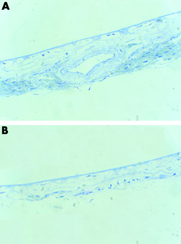
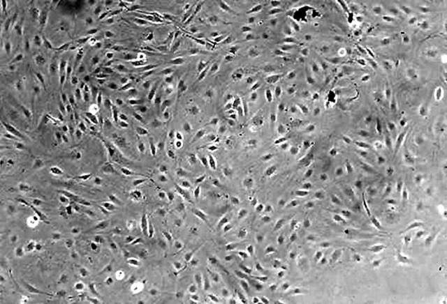
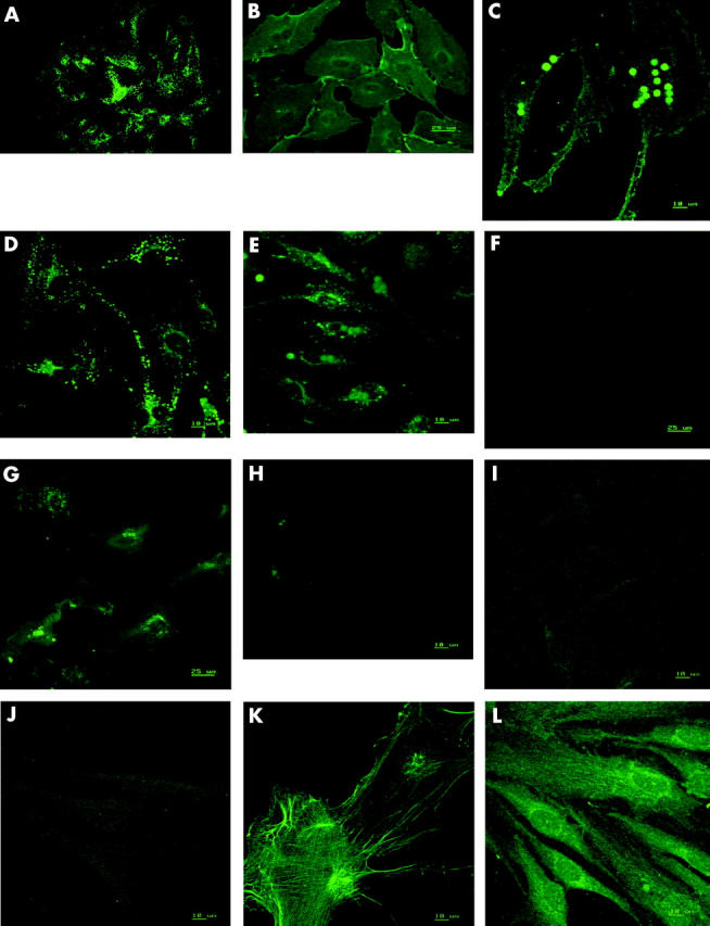
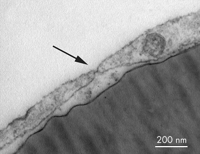
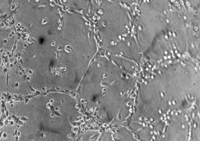
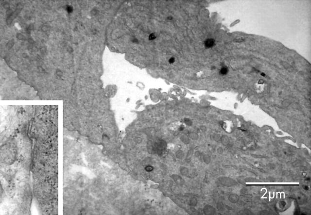
Method: Human ICECs were isolated after manual dissection of maculas from fresh human posterior segments. Following enzyme digestion to form a single cell suspension, the ICECs were isolated using anti-CD31 coated Dynabeads. The isolated cells were grown in culture and examined for typical endothelial cell morphology, surface expression of vWf, CD 31, CD 105, VEGF receptors 1 and 2, and expression of E-selectin after stimulation with TNF-alpha. The cells were also examined for their ability to form fenestrations and capillary-like tubes in Matrigel.
Results: The method enabled the rapid isolation of viable cells that demonstrated typical endothelial cobblestone morphology in culture. The cells stained positive for CD31, vWf, CD105, VEGF receptors 1 and 2, and E-selectin (after stimulation with TNF-alpha). The cells stained negative for alpha smooth muscle actin and fibroblast surface protein. The cells also developed fenestrations when cultured on fibronectin coated plates and formed capillary-like tubes structures when cultured on Matrigel.
Conclusions: This technique isolates cells from the human macular inner choroid that display features consistent with vascular endothelial cells. These cells could subsequently be used to further the understanding of the pathophysiological mechanisms of diseases of the inner choroid, such as choroidal neovascularisation.
Figures
References
-
- Gass JDM. Biomicroscopic and histopathologic considerations regarding the feasibility of surgical excision of subfoveal neovascular membranes. Am J Ophthalmol 1994;118:285–98. - PubMed
-
- Sarks JP, Sarks SH, Killingsworth MC. Morphology of early choroidal neovascularisation in age-related macular degeneration: correlation with activity. Eye 1997;11:515–22. - PubMed
-
- Killingworth MC. Angiogenesis in early choroidal neovascularisation secondary to age-related macular degeneration. Graefes Arch Clin Exp Ophthalmol 1995;233:313–23. - PubMed
-
- Liu X, Ye X, Yanoff M, et al. Regulatory effects of soluble growth factors on choriocapillaris endothelial growth and survival. Ophthalmic Res 1998;30:302–13. - PubMed
-
- Aird WC. Endothelial cell heterogeneity. Crit Care Med 2003;31:S221–S230. - PubMed
Publication types
MeSH terms
Substances
LinkOut - more resources
Full Text Sources
Miscellaneous