Contribution to comprehension of image formation in confocal microscopy of cornea with Rostock cornea module
- PMID: 16170131
- PMCID: PMC1772867
- DOI: 10.1136/bjo.2004.063743
Contribution to comprehension of image formation in confocal microscopy of cornea with Rostock cornea module
Abstract
Aim: To investigate the influence of refractive index of aqueous humour on imaging of corneal endothelium in confocal microscopy. To clarify the phenomenon of dark endothelial and bright epithelial cell membranes in confocal images of corneas.
Methods: Use of a novel digital confocal laser scanning microscope, a combination of the Heidelberg retina tomograph (HRT II) and the Rostock cornea module. Exchange of aqueous humour solution from domestic pigs against glycerol/water solutions (refractive indices eta = 1.337-1.47). Transelectron microscopy of endothelial and epithelial cell morphology.
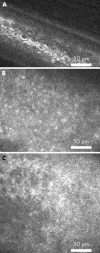
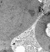
Results: Under the terms of variable refractive indices no differences were observed for general imaging of endothelium. Bright cells were bordered by dark cell membranes in all experiments. Electron microscopy of endothelium and epithelium revealed differences in intracellular and cell membrane structure of both cell types.
Conclusion: Source of specific confocal optical behaviour of endothelium does not come from interface conditions to aqueous humour, but may result from intracellular variations and ultrastructure of cell membranes.
Figures


References
-
- Calvillo MP, McLaren JW, Hodge DO, et al. Corneal reinnervation after LASIK: prospective 3-year longitudinal study. Invest Ophthalmol Vis Sci 2004;45:3991–6. - PubMed
-
- Nakano E, Oliveira M, Portellinha W, et al. Confocal microscopy in early diagnosis of Acanthamoeba keratitis. J Refract Surg 2004;20 (Suppl) :737–40. - PubMed
-
- Patel S, McLaren J, Hodge D, et al. Normal human keratocyte density and corneal thickness measurement by using confocal microscopy in vivo. Invest Ophthalmol Vis Sci 2001;42:333–9. - PubMed
-
- Guell JL, Velasco F, Guerrero E, et al. Confocal microscopy of corneas with an intracorneal lens for hyperopia. J Refract Surg 2004;20:778–82. - PubMed
-
- Mastropasqua L, Nubile M. Confocal microscopy of the cornea. Thorofare, NJ: Slack, 2002.
MeSH terms
Substances
LinkOut - more resources
Full Text Sources
Research Materials
Miscellaneous
