. 2005 Nov 18;280(46):38464-70.
doi: 10.1074/jbc.M508744200. Epub 2005 Sep 16.

The glycolytic enzymes, glyceraldehyde-3-phosphate dehydrogenase, triose-phosphate isomerase, and pyruvate kinase are components of the K(ATP) channel macromolecular complex and regulate its function

Affiliations

The glycolytic enzymes, glyceraldehyde-3-phosphate dehydrogenase, triose-phosphate isomerase, and pyruvate kinase are components of the K(ATP) channel macromolecular complex and regulate its function

Piyali Dhar-Chowdhury et al. J Biol Chem. .

Abstract

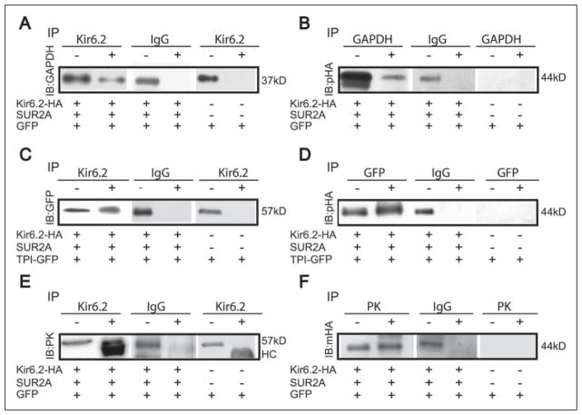
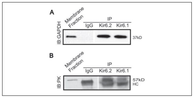
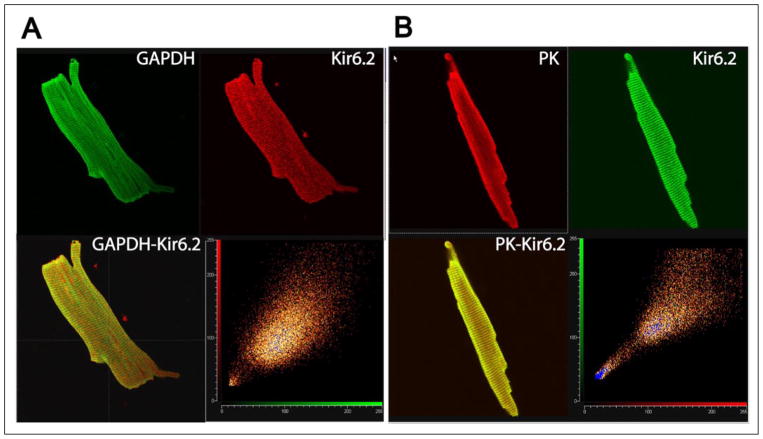
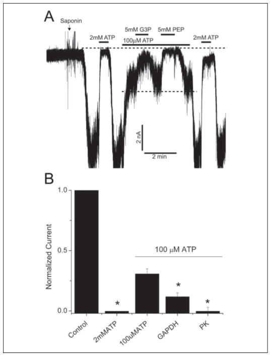
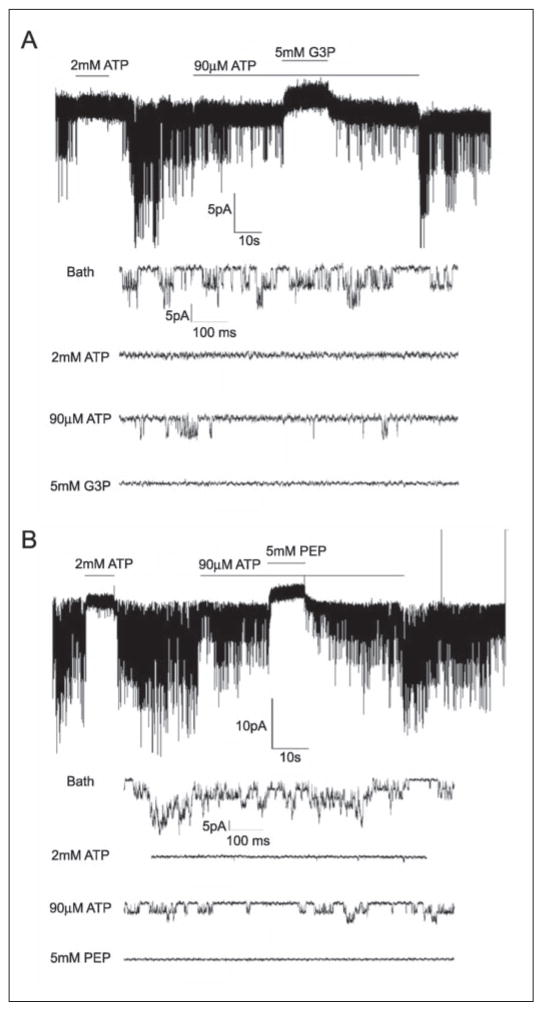
The regulation of ATP-sensitive potassium (K(ATP)) channel activity is complex and a multitude of factors determine their open probability. Physiologically and pathophysiologically, the most important of these are intracellular nucleotides, with a long-recognized role for glycolytically derived ATP in regulating channel activity. To identify novel regulatory subunits of the K(ATP) channel complex, we performed a two-hybrid protein-protein interaction screen, using as bait the mouse Kir6.2 C terminus. Screening a rat heart cDNA library, we identified two potential interacting proteins to be the glycolytic enzymes, glyceraldehyde-3-phosphate dehydrogenase (GAPDH) and triose-phosphate isomerase. The veracity of interaction was verified by co-immunoprecipitation techniques in transfected mammalian cells. We additionally demonstrated that pyruvate kinase also interacts with Kir6.2 subunits. The physiological relevance of these interactions is illustrated by the demonstration that native Kir6.2 protein similarly interact with GAPDH and pyruvate kinase in rat heart membrane fractions and that Kir6.2 protein co-localize with these glycolytic enzymes in rat ventricular myocytes. The functional relevance of our findings is demonstrated by the ability of GAPDH or pyruvate kinase substrates to directly block the K(ATP) channel under patch clamp recording conditions. Taken together, our data provide direct evidence for the concept that key enzymes involved in glycolytic ATP production are part of a multisubunit K(ATP) channel protein complex. Our data are consistent with the concept that the activity of these enzymes (possibly by ATP formation in the immediate intracellular microenvironment of this macromolecular K(ATP) channel complex) causes channel closure.


Figures

FIGURE 1
FIGURE 1. Kir6.2 subunits interact with GAPDH, TPI, and PK in transfected cells
Lysates of COS7L cells were transfected with Kir6.2-HA and SUR2A, as indicated at the bottom of each blot (GFP was used as a marker of transfection efficiency). Some cells were additionally co-transfected with TPI-GFP cDNA. Kir6.2 subunits were immunoprecipitated (IP) with anti-Kir6.2 (W62) antibody. Other precipitating antibodies used were anti-GAPDH, anti-PK, and anti-GFP antibodies. Immunoprecipitates were subjected to SDS-PAGE and immunoblotting was performed using anti-GAPDH, monoclonal anti-HA, polyclonal anti-HA, monoclonal anti-GFP, or anti-PK antibodies. As a negative control, IP reactions were performed with untransfected COS7L cells or IgG antibodies. The first lane of each immunoblot (−) is a sample of the cell lysate without an immunoprecipitation reaction to demonstrate that the relevant protein is expressed in the cells. The second lane (+) is obtained from the immunoprecipitate, using the antibodies as indicated. The approximate molecular sizes are indicated (HC, IgG heavy chain).
FIGURE 2
FIGURE 2. Native Kir6.2 and Kir6.1 subunits associate with GAPDH and PK in rat heart membranes
Rat heart membrane fractions were used in immunoprecipitating (IP) reactions with anti-Kir6.2 antibodies (W62) or Kir6.1 antibodies (NAF1). The immuno-precipitates were separated on 12% SDS-PAGE and the resulting immunoblots (IB) were probed using anti-GAPDH antibody (A) or anti-PK antibody (B). The positive control for immunoblotting was the cardiac membrane fraction labeled membrane fraction (lane 1 in each case). GAPDH (37 kDa) and PK (57 kDa) were both detected in the Kir6.2 and Kir6.1 immunoprecipitates. Co-immunoprecipitation with IgG antibodies as a negative control failed to detect either GAPDH or PK. Because of cross-reactivity between the polyclonal antibodies used in panel B, IgG heavy chain IgG is observed at ~50 kDa (denoted HC). TPI could not be detected in native tissue because of lack of commercial antibodies.
FIGURE 3
FIGURE 3. Kir6.2 subunits colocalize with GAPDH and PK in rat ventricular cardiomyocytes
A, myocytes were double stained with anti-Kir6.2 and anti-GAPDH antibodies (top panel, secondary antibodies, respectively, were Cy-3 conjugated donkey anti-rabbit IgG, red; and Cy-2 conjugated donkey anti-mouse IgG, green). The confocal image was acquired halfway through the height of the myocyte in the Z-direction. B, cardiomyocytes were double stained with anti-Kir6.2 and anti-PK antibodies. The secondary antibodies used were Cy5-conjugated donkey anti-rabbit IgG (pseudo colored green) and Cy3-conjugated donkey anti-goat IgG (pseudo-colored red). Yellow depicts co-localization. The bottom right panel is a scatter plot of corresponding pixel intensities of the green and red channel.
FIGURE 4
FIGURE 4. GAPDH and PK substrates inhibit rat ventricular KATP channels in the open cell-configuration
A, a representative trace showing the effects of GAPDH and PK substrates on KATP channel activity in rat ventricular myocytes. The intracellular bath solution contained 100 μM ATP to prevent rigor contracture of the myocytes. Substances were added as indicated by the horizontal bars. The dotted lines, respectively, depict the closed state of the channel and the partially blocked state in the presence of 100 μM ATP, 0.5 mM ADP, and 0.5 mM KH2PO4 (depicted as 100 μM ATP for simplicity). The patch potential was −60 mV (pipette potential of +60 mV). The current was filtered at 1 kHz. B, averages of mean patch KATP channel current (mean ± S.E.), normalized to mean patch current in the absence of ATP, under the various conditions, as indicated. n = 6, *, p < 0.05 compared with the 100 μM ATP group. G3P, glyceraldehyde 3-phosphate.
FIGURE 5
FIGURE 5. Substrates of GAPDH and PK suppress KATP channel in excised, inside-out membrane patches
The bath and pipette solutions were essentially the same as in the previous figure. A, a representative trace showing the effect of GAPDH substrates. Channel activity was blocked by 2 mM ATP and partially inhibited by 90 μM ATP (with 0.5 mM ADP and 0.5 mM KH2PO4). Further addition of glyceraldehyde 3-phosphate (G3P) (5 mM; also containing 1 mM NADP) reversibly inhibited channel activity. Expanded traces below show channel activity under selected conditions at a faster time scale. B, PK substrates (5 mM PEP) also inhibited KATP current. The format is the same as in the previous panel.

References

    1. Seino S, Miki T. Prog Biophys Mol Biol. 2003;81:133–176. - PubMed
    1. Inagaki N, Seino S. Jpn J Physiol. 1998;48:397–412. - PubMed
    1. Leonoudakis D, Conti LR, Anderson S, Radeke CM, McGuire LM, Adams ME, Froehner SC, Yates JR, III, Vandenberg CA. J Biol Chem. 2004;279:22331–22346. - PubMed
    1. Leonoudakis D, Conti LR, Radeke CM, McGuire LM, Vandenberg CA. J Biol Chem. 2004;279:19051–19063. - PubMed
    1. Bers DM. J Mol Cell Cardiol. 2004;37:417–429. - PubMed

Publication types

MeSH terms

LinkOut - more resources