

Review
doi: 10.1055/s-2005-917662.
Neuroparasitic infections: nematodes
Affiliations
- PMID: 16170738
- PMCID: PMC2678030
- DOI: 10.1055/s-2005-917662
Item in Clipboard
Review
Neuroparasitic infections: nematodes
Semin Neurol.
2005 Sep.
Abstract
Globalization has produced an increase in the number of people at risk for contracting parasitic infection. Central nervous system infection by nematodal parasites can be devastating. Early recognition and treatment of infection can significantly decrease morbidity of the parasitic infection, as well as the risk of secondary superinfection. The clinical presentation, diagnosis, and treatment for five of the more common nematodal infections of the nervous system--Angiostrongylus spp., Baylisacaris procyonis, Gnathostoma spinigerum, Strongyloides stercoralis, and Toxocara spp.--is reviewed.
Figures
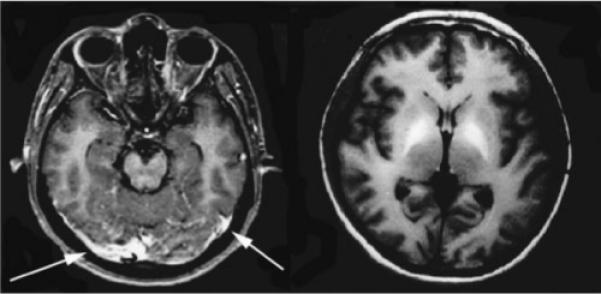
Patient with Angiostrongylus infection. Axial T1 contrast-enhanced images demonstrate meningeal enhancement (left, arrows) and markedly increased signal intensity within the globus pallidus (right). (Reprinted with permission from Tsai HC, Liu YC, Kunin CM, et al. Eosinophilic meningitis caused by Angiostrongylus cantonensis associated with eating raw snails: correlation of brain magnetic resonance imaging scans with clinical findings. Am J Trop Med Hyg 2003;68:281−285.)
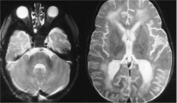
Thirteen month-old patient with Baylisascaris infection. Bilateral, patchy T2 hyperintensity is seen predominantly in the white matter, including the periventricular regions and corpus medullaris of the cerebellum. (Images courtesy of Dr. Howard Rowley.)
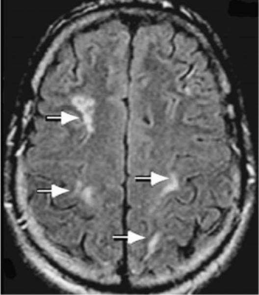
Patient with Gnathostoma infection. Axial FLAIR MRI of the brain showing bilateral clusters of small rounded hyperintensities in deep white matter (arrows). (Reprinted with permission from Hughes AJ, Biggs BA. Parasitic worms of the central nervous system: an Australian perspective. Intern Med J 2002;32:541−553.)
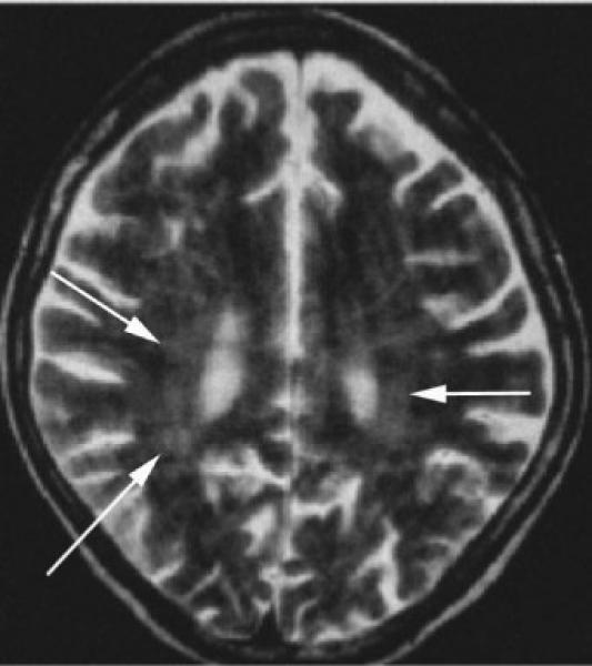
Thirty-five-year-old immunosuppressed patient with Strongyloides infection. Axial T2-weighted images reveal global atrophy and patchy periventricular white matter hyperintensities (arrows). (Reprinted with permission from Kothary NN, Muskie JM, Mathur SC. Strongyloides stercoralis hyperinfection. Radio-graphics 1999;19:1077−1081.)
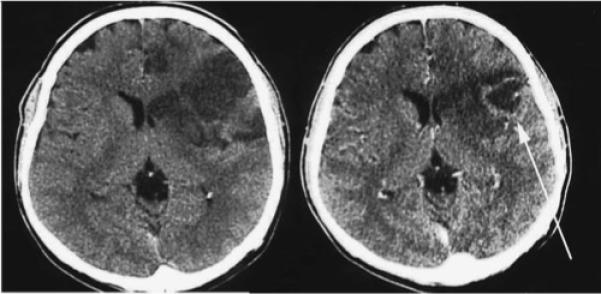
Patient with Toxocara infection. Noncontrast CT imaging demonstrates left frontal hypodensity consistent with edema (left), and contrast-enhanced imaging reveals a ring enhancing lesion (arrow on right). (Reprinted with permission from Oktar N, Barçin E, Kazandi A, Korkmaz M. Cerebral Toxocara mimicking a malignant glioma. Norol Bil D 2002;19:#12.)
References
-
- Jindrak K. Angiostrongyliasis cantonensis (eosinophilic meningitis, Alicata's disease). Contemp Neurol Ser. 1975;12:133–164. - PubMed
-
- Chau TT, Thwaites GE, Chuong LV, et al. Headache and confusion: the dangers of a raw snail supper. Lancet. 2003;361:1866. - PubMed
-
- Andersen E, Gubler DJ, Sorensen K, et al. First report of Angiostrongylus cantonensis in Puerto Rico. Am J Trop Med Hyg. 1986;35:319–322. - PubMed
-
- Slom TJ, Cortese MM, Gerber SI, et al. An outbreak of eosinophilic meningitis caused by Angiostrongylus cantonensis in travelers returning from the Caribbean. N Engl J Med. 2002;346:668–675. - PubMed
Publication types
MeSH terms
Grants and funding
LinkOut - more resources
Full Text Sources
Other Literature Sources
Medical
Miscellaneous
