Comparative Study
. 2005 Nov 30;141(2):138-50.
doi: 10.1016/j.molbrainres.2005.08.011. Epub 2005 Sep 19.

Environmental enrichment increases progenitor cell survival in the dentate gyrus following lateral fluid percussion injury

Affiliations
Comparative Study

Environmental enrichment increases progenitor cell survival in the dentate gyrus following lateral fluid percussion injury

Lindsey J Gaulke et al. Brain Res Mol Brain Res. .

Abstract

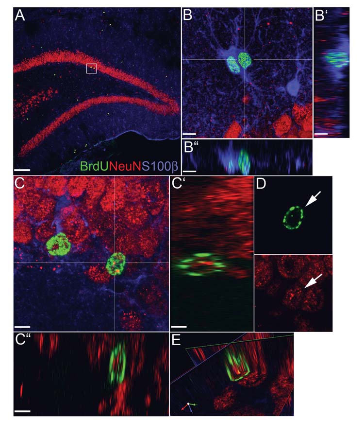
Neurons in the hilus of the dentate gyrus are lost following a lateral fluid percussion injury. Environmental enrichment is known to increase neurogenesis in the dentate in intact rats, suggesting that it might also do so following fluid percussion injury, and potentially provide replacements for lost neurons. We report that 1 h of daily environmental enrichment for 3 weeks increased the number of progenitor cells in the dentate following fluid percussion injury, but only on the ipsilesional side. In the dentate granule cell layer, but not the hilus, most progenitors had a neuronal phenotype. The rate of on going cell proliferation was similar across groups. Collectively, these results suggest that the beneficial effects of environmental enrichment on behavioral recovery following FP injury are not attributable to neuronal replacement in the hilus but may be related to increased neurogenesis in the granule cell layer.


Figures

Figure 1
Figure 1
Immunofluorescent images produced from captured digital confocal laser scanning microscopy illustrating dividing cells (BrdU, green), mature neurons (NeuN, red), and astrocytes (S100β, blue) in the dentate granule cell layer and hilus 3 weeks following a FP injury (A). Cells that are co-labeled with BrdU and S100b are found throughout the dentate and hilar structures (B). A merged confocal z-stack illustrates colocalization of S100β and BrdU within the nucleus of two cells (B). The cytoplasmic compartment is clearly enveloped by S100β staining shown in orthogonal views (B’,B” taken at point of intersecting lines in B). NeuN and BrdU double-labeled cells are found within the dentate gyrus of all injured rodents. A high power image taken from highlighted region in A shows 2 BrdU and NeuN colabeled cells (C). Orthogonal views taken from the center of one neuron demonstrates good correlation but limited overlap of BrdU and NeuN signals within the nuclear compartment (C’,C”). Individual 1μm-thick confocal planes show the neuronal morphology and separation of the green and red signals within the BrdU-labeled neuron (D). A 3-D reconstruction of a labeled cell taken from the cross-hairs in C and rotated at the center of the BrdU-labeled nucleus (E). Scale Bars: (A=70μm), (B, B’, B”=4.5μm), (C, C’, C”=3.5 μm)
Figure 2
Figure 2
Effect of EE on cell proliferation in the dentate following FP injury. Schematic representation of BrdU+ cells (filled circles) in the granule cell layer (g.c.) and hilus of the dentate gyrus from representative animals following sham injury (A), FP injury (B), and FP injury + EE (C). Digital images were acquired using the FITC and TRITC lasers of a confocal microscope, merged, and then imported into Photoshop. A montage of the images was used into to prepare a diagram of the entire dentate gyrus, following which, the borders of the granule cell layer and CA3 regions were outlined, and the location of BrdU+ cells were marked in the hilus and granule cell layer. The graph (D) depicts numbers of BrdU+ cells in the dentate gyrus by subregions: left granule cell layer and subgranular zone (LGC); right (RGC); left hilus (L HIL) and right (R HIL), 3 weeks following a FP injury (FP), FP injury + environmental enrichment (FP+EE), or a sham injury (SH). FP+EE increased total BrdU+ cells in the LGC compared to FP (p < 0.002) and SH (p < 0.001), and in the L HIL compared to SH (p < 0.02). Scale bars equal 100 μm. *Indicates significant increases in comparison to one or both of the other groups.
Figure 3
Figure 3
Effects of EE on cell fate in the dentate following FP. Graphs depict numbers (A,C,E) and percentages (B,D,F) of double-labeled and single-labeled BrdU cells by subregions of the dentate gyrus. Double-labeled BrdU/NeuN cells were significantly increased in the LGC for FP+EE compared to FP (p < 0.003) and SH (p < 0.001) (A). The percentage of double-labeled BrdU/NeuN cells in the L granule cell layer, though, was actually greater for SH compared to FP and FP+EE (p < 0.02 and 0.05, respectively) (B). The number and percentage of double-labeled BrdU/S100β cells did not differ between FP and FP+EE, but were increased in comparison to the SH group (p < 0.01 and 0.001, respectively) (C, D). Numbers of single-labeled BrdU cells for FP+EE were also increased in the LGC compared to SH (p < 0.05) (E). In the L hilus, there was a significant increase in single-labeled BrdU cells compared to FP (p < 0.03) and to SH (p < 0.01) (E). *Indicates significant increases in comparison to one or both of the other groups.
Figure 4
Figure 4
Effects of rostral-caudal level on cell proliferation and fate following FP or FP+EE compared to SH. Graphs depict numbers of single- and double-labeled BrdU cells at various bregma levels for the left granule cell layer (A, B) or hilus (C, D) for each group. A repeated measures ANOVA demonstrated that in the left granule cell layer and hilus the number of BrdU/Sl00B cells varied significantly by bregma level (p < 0.002). In contrast to the effects of rostral-caudal location on gliogenesis, no differences were observed for neurogenesis (B) or single-labeled cells (D).
Fig. 5
Fig. 5
Effect of EE on chronic cell proliferation in the dentate gyrus following FP. Photomicrograph depicts Ki67+ cells in a coronal section of the granule cell layer (g.c.) and hilus and of the dentate gyrus 3 weeks following a FP injury (A). Most, but not all, of the labeled cells were in the subgranular zone. Inset is higher magnification of cells in the subgranular zone. Graph depicts numbers of Ki67+ cells in the right and left granule cell layers and hilar regions. Numbers did not significantly vary between groups, although there was a trend for a decrease in the FP+EE group (B). Scale bar = 50 μm.
Figure 6
Figure 6
EE attenuates cognitive deficits after FP injury. Graph depicts MWM goal latencies (group means) for each trial block. Animals were given 2 trial blocks (4 trials each) per day for 2 days. Following a repeated measures ANOVA, post hoc tests indicated that the FP+EE and SH performed significantly better than FP (p < 0.001).

References

    1. Ambrogini P, Cuppini R, Cuppini C, Ciaroni S, Cecchini T, Ferri P, Sartini S, Del Grande P. Spatial learning affects immature granule cell survival in adult rat dentate gyrus. Neurosci Lett. 2000;286:21–4. - PubMed
    1. Arvidsson A, Collin T, Kirik D, Kokaia Z, Lindvall O. Neuronal replacement from endogenous precursors in the adult brain after stroke. Nat Med. 2002;8:963–70. - PubMed
    1. Baldwin SA, Scheff SW. Intermediate filament change in astrocytes following mild cortical contusion. Glia. 1996;16:266–75. - PubMed
    1. Bramlett HM, Dietrich WD. Quantitative structural changes in white and gray matter 1 year following traumatic brain injury in rats. Acta Neuropathol (Berl) 2002;103:607–14. - PubMed
    1. Briones TL, Suh E, Hattar H, Wadowska M. Dentate gyrus neurogenesis after cerebral ischemia and behavioral training. Biol Res Nurs. 2005;6:167–79. - PubMed

Publication types

MeSH terms