. 2005 Sep 26;170(7):1047-55.
doi: 10.1083/jcb.200503023. Epub 2005 Sep 19.

Aurora A phosphorylation of TACC3/maskin is required for centrosome-dependent microtubule assembly in mitosis

Affiliations

Aurora A phosphorylation of TACC3/maskin is required for centrosome-dependent microtubule assembly in mitosis

Kazuhisa Kinoshita et al. J Cell Biol. .

Abstract

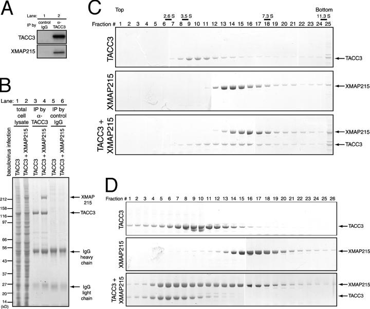
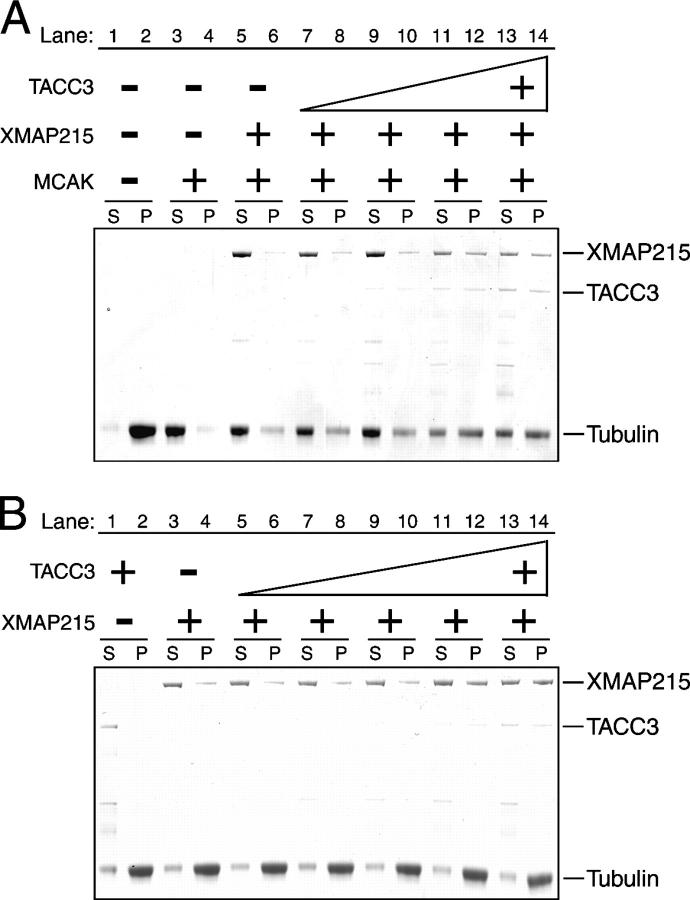
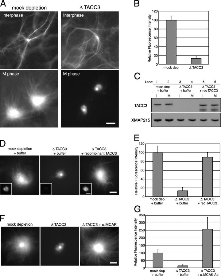
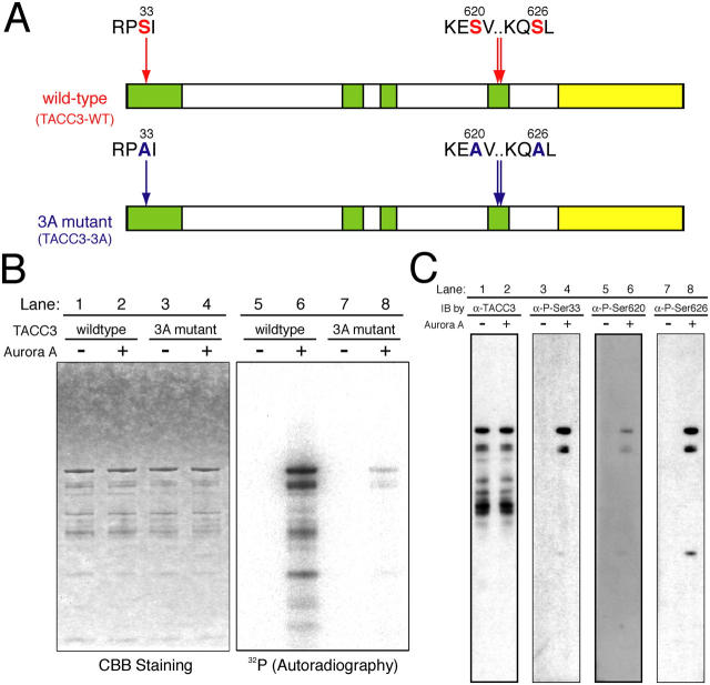
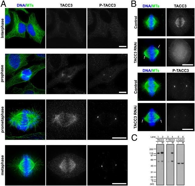
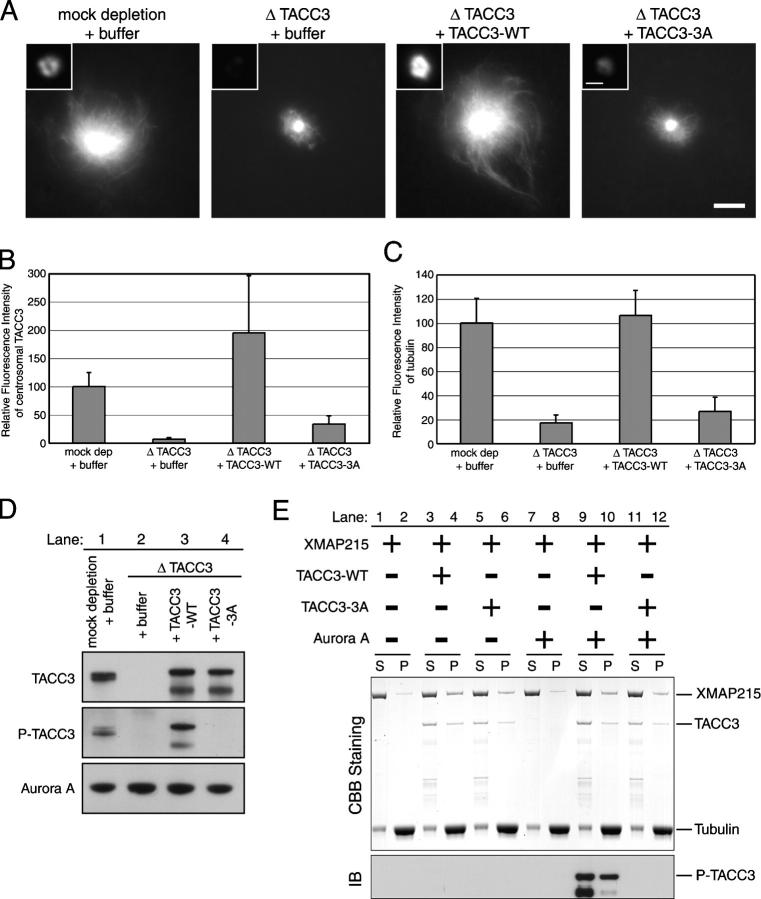
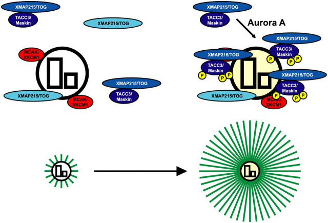
Centrosomes act as sites of microtubule growth, but little is known about how the number and stability of microtubules emanating from a centrosome are controlled during the cell cycle. We studied the role of the TACC3-XMAP215 complex in this process by using purified proteins and Xenopus laevis egg extracts. We show that TACC3 forms a one-to-one complex with and enhances the microtubule-stabilizing activity of XMAP215 in vitro. TACC3 enhances the number of microtubules emanating from mitotic centrosomes, and its targeting to centrosomes is regulated by Aurora A-dependent phosphorylation. We propose that Aurora A regulation of TACC3 activity defines a centrosome-specific mechanism for regulation of microtubule polymerization in mitosis.


Figures

Figure 1.
Figure 1.
TACC3 forms a complex with XMAP215 in Xenopus egg extracts and in vitro. (A) Coimmunoprecipitation in Xenopus egg extracts. Immunoprecipitation (IP) was performed using control IgG (lane 1) or anti-TACC3 antibody (lane 2). The blots were probed with anti-TACC3 (top) or anti-XMAP215 (bottom). (B) Coimmunoprecipitation in the extracts of baculovirus-infected insect cells. Total lysates of TACC3 baculovirus single infected cells and TACC3 (lane 1) and XMAP215 virus double infected cells (lane 2) were prepared for immunoprecipitation. In each cell lysate, immunoprecipitation were performed using anti-TACC3 (lanes 3 and 4) or control IgG antibody (lanes 5 and 6). Total cell lysate and immunoprecipitates that were dissolved in sample buffer were subjected to SDS-PAGE, and the gel was stained with Coomassie brilliant blue. (C) Fractions from the top and bottom of a continuous sucrose density gradient centrifugation with purified proteins. Fractions collected after 3–15% continuous sucrose gradient centrifugation with TACC3 alone (top), XMAP215 alone (middle), or a mixture of TACC3 and XMAP215 (bottom) were subjected to SDS-PAGE. The gels were stained with Coomassie brilliant blue. (D) Gel filtration with purified proteins. TACC3 alone (top), XMAP215 alone (middle), or a mixture of TACC3 and XMAP215 (bottom) were analyzed by gel filtration. Fractions were collected and subjected to SDS-PAGE, and the gels were stained with Coomassie brilliant blue.
Figure 2.
Figure 2.
TACC3 increases the antagonizing activity of XMAP215 against MCAK. (A) Sedimentation analysis to monitor MCAK-dependent microtubule destabilization activity. 2.5 μM GMPCPP microtubules were mixed with 125 nM MCAK (lanes 3–14) or control buffer (lanes 1 and 2) in presence of ATP. 500 nM XMAP215 (lanes 5–14) and increasing concentrations of TACC3 from 250 (lanes 7 and 8), 500 (9 and 10), and 1,000 (11 and 12) to 1,500 nM (13 and 14) or control buffer (5 and 6) were added in reactions. Reaction mixtures were sedimented after a 38-min incubation at 30°C, supernatants (S) and pellets (P) were subjected to SDS-PAGE, and the gel was stained with Coomassie brilliant blue. (B) Sedimentation assay to monitor XMAP215 affinity to microtubules. 2.5 μM GMPCPP microtubules were mixed with 1,250 nM TACC3 (lanes 1 and 2), 500 nM XMAP215 (3 and 4), or 500 nM XMAP215 in addition to increasing concentrations of TACC3 from 250 (5 and 6), 500 (7 and 8), 750 (9 and 10), and 1,000 (11 and 12) to 1,250 nM (13 and 14). Mixtures were incubated for 38 min at 30°C and sedimented, and supernatants (S) and pellets (P) were analyzed by SDS-PAGE.
Figure 3.
Figure 3.
TACC3 is required for mitosis-specific microtubule assembly around centrosomes. (A) Microtubules were visualized by the addition of fluorescently labeled tubulin in control mock-depleted (left) and TACC3-depleted extracts (right). Centrosomes were added to interphase extracts (top), and then the extracts were driven into M phase by the addition of nondegradable cyclin B (bottom). Bar, 10 μm. (B) Quantification of tubulin fluorescence in A. (C) Immunoblots of depleted extracts. The extracts were immunodepleted with control IgG (lanes 1 and 2; mock dep) or anti-TACC3 antibody (lanes 3–6; Δ TACC3) with adding back of TACC3 (lanes 5 and 6) or control buffer (lanes 1–4). The blots of interphase (lanes 1, 3, and 5) and mitotic extracts (lanes 2, 4, and 6) were probed by anti-TACC3 (top; TACC3) and anti-XMAP215 (bottom; XMAP215). (D) Microtubules assembled around mitotic centrosomes in immunodepleted extracts with add back of recombinant proteins. Purified recombinant TACC3 or control buffer were added in mock-depleted or TACC3-depleted extracts. Bar, 10 μm. Insets show immunolocalization of TACC3 on centrosomes. Bar, 1 μm. (E) Quantification of tubulin fluorescence in D. (F) Inhibition of MCAK in TACC3-depleted extracts. Microtubules were assembled around centrosomes in mock-depleted extracts plus control buffer (left), TACC3-depleted extracts plus control buffer (middle), or anti-MCAK antibody (right). The addition of inhibitory MCAK antibody rescued the effect of TACC3 depletion on microtubule assembly around mitotic centrosomes. Bar, 10 μm. (G) Quantification of tubulin fluorescence in F. (B, E, and G) Relative fluorescence intensity of labeled tubulin around mitotic centrosomes (within 21.079 μm radius) was measured. Values shown are the means plus SD.
Figure 4.
Figure 4.
Determination of Aurora A phosphorylation sites of TACC3 in vitro . (A) Consensus sequences for Aurora A phosphorylation in Xenopus TACC3/maskin. Yellow boxes indicate conserved domains among the TACC family, whereas green boxes are the domains that are highly conserved with TACC3 homologues only. The 3A mutant protein (TACC3-3A) has mutations of alanine substitution on three serine residues (Ser33, Ser620, and Ser626) in the consensus sequences of Aurora A phosphorylation. Numbers represent the positions for amino acid residues of Aurora A phosphorylation target sites in the amino acid sequence of TACC3. (B) Aurora A kinase assay with recombinant WT versus alanine mutant TACC3 proteins. WT or 3A mutant TACC3 was incubated with or without recombinant Aurora A (−/+ Aurora A) in the presence of γ−[32P] ATP (see Materials and methods). The reaction mixture was loaded onto SDS-PAGE, and the gel was stained with Coomassie brilliant blue (CBB; lanes 1–4). The incorporation of 32P into TACC3 in the gel was measured by autoradiography (32P; lanes 5–8). (C) Characterization of phosphospecific antibodies. TACC3-WT was incubated with or without Aurora A (−/+ Aurora A), and the reaction mixture was loaded onto SDS-PAGE. The blots were probed with anti-TACC3 antibody (lanes 1 and 2), antiphospho-Ser33 (lanes 3 and 4), antiphospho-Ser620 (lanes 5 and 6), and antiphospho-Ser626 (lanes 7 and 8).
Figure 5.
Figure 5.
Aurora A phosphorylated TACC3 is enriched at mitotic centrosomes. (A) Immunolocalization of TACC3 and phospho-TACC3. Deconvolved images of human tissue culture cells stained for DNA (blue)/microtubules (MTs; green), TACC3, and phospho-TACC3 (P-TACC3; stained by antiphospho-Ser626 antibodies) in different cell cycle stages. Bars, 10 μm. (B) Immunolocalization of TACC3 and phospho-TACC3 in siRNA-treated cells. Deconvolved images of either control or TACC3 RNA interference–treated cells stained for DNA (blue)/microtubules (MTs; green), TACC3 (top), and phospho-TACC3 (P-TACC3; bottom). Arrows indicate misaligned chromosomes that are indicative of the TACC3 RNA interference phenotype. Bar, 10 μm. (C) Phosphorylation of TACC3 is mitosis specific in human tissue culture cells. The TACC3 antibody detects a protein of ∼130 kD in extracts prepared from either an asynchronous culture of HeLa S3 cells (lane 1; I) or cells arrested in mitosis by nocodazole treatment (lane 2; M). The phosphospecific TACC3 antibody (antiphospho-Ser626) recognizes a band with the same molecular mass size in mitotic cells (lane 4) but not in asynchronously cultured cells (lane 3). The blot probed by α-tubulin antibody is a loading control (lane 5, asynchronous cultured cells; lane 6, mitotic arrested cells).
Figure 6.
Figure 6.
Phosphorylation of TACC3 by Aurora A is required for centrosomal localization of TACC3 and its microtubule-stimulating activity. (A) Localization of WT versus phosphorylation mutant of TACC3 on mitotic centrosomes. Microtubules around mitotic centrosomes were visualized by the addition of fluorescent tubulin in add-back experiments. Bar, 10 μm. Insets show immunolocalization of TACC3 on centrosomes in immunodepleted extracts with add back of WT versus the phosphorylation mutant of TACC3. Bar, 1 μm. (B) Quantification of immunostaining of TACC3 on centrosomes in A. Relative fluorescence intensity of TACC3 staining on centrosomes (within 1.079 μm radius) was measured. (C) Quantification of tubulin fluorescence in A. Relative fluorescence intensity of labeled tubulin around mitotic centrosomes (within 21.079 μm radius) was measured. (B and C) Values shown are the means plus SD. (D) Immunoblots of depleted extracts in add-back experiments. Mitotic extracts were immunodepleted with control IgG (lane 1; mock dep) or anti-TACC3 antibody (lanes 2–4; Δ TACC3) with adding back of control buffer (lanes 1 and 2; + buffer), TACC3-WT (lane 3; + TACC3-WT), or phosphorylation mutant TACC3 (lanes 4; + TACC3-3A). (E) Sedimentation assay to monitor XMAP215 affinity to microtubules in the presence of WT or phosphorylation mutant TACC3. 2.5 μM GMPCPP microtubules were mixed with 0.5 μM XMAP215 alone (lanes 1, 2, 7, and 8), 0.5 μM XMAP215 + 1.0 μm TACC3-WT (lanes 3, 4, 9, and 10), or 0.5 μM XMAP215 + 1.0 μm TACC3-3A (lanes 5, 6, 11, and 12) in the absence (lanes 1–6) or presence (lanes 7–12) of Aurora A. Mixtures were incubated for 38 min at 30°C and sedimented, and supernatants (S) and pellets (P) were subjected to SDS-PAGE. The gels were stained with Coomassie brilliant blue (top; CBB staining) or transferred onto nitrocellurose membrane for immunoblotting (IB) using the phosphospecific TACC3 antibody (antiphospho-Ser626; bottom).
Figure 7.
Figure 7.
A model of centrosomal microtubule assembly in M phase. XKCM1/MCAK (red) localized on centrosomes generates an environment in which plus end growth of microtubules (green) is not favored, and XMAP215/TOG (light blue) cannot stabilize nascent nucleated plus ends (left). Aurora A phosphorylation of TACC3/maskin targets the TACC3–XMAP215 complex (dark blue) to centrosomes. The targeting enhances the activity of XMAP215 at centrosomes, stabilizing nascent plus ends and allowing microtubules to grow from centrosomes (right). The encircled P (yellow) represents the serine residues that were phosphorylated by Aurora A in TACC3.

Similar articles

Cited by

References

    1. Ashford, A.J., S.S. Andersen, and A.A. Hyman. 1998. Preparation of tubulin from bovine brain. Cell Biology: A Laboratory Handbook. Vol. 2. 2nd ed. J.E. Celis, editor. Academic Press/UK, London. 205–212.
    1. Bellanger, J.M., and P. Gönczy. 2003. TAC-1 and ZYG-9 form a complex that promotes microtubule assembly in C. elegans embryos. Curr. Biol. 13:1488–1498. - PubMed
    1. Belmont, L.D., A.A. Hyman, K.E. Sawin, and T.J. Mitchison. 1990. Real-time visualization of cell cycle-dependent changes in microtubule dynamics in cytoplasmic extracts. Cell. 62:579–589. - PubMed
    1. Bischoff, J.R., L. Anderson, Y. Zhu, K. Mossie, L. Ng, B. Souza, B. Schryver, P. Flanagan, F. Clairvoyant, C. Ginther, et al. 1998. A homologue of Drosophila aurora kinase is oncogenic and amplified in human colorectal cancers. EMBO J. 17:3052–3065. - PMC - PubMed
    1. Cheeseman, I.M., S. Anderson, M. Jwa, E.M. Green, J. Kang, J.R. Yates, C.S. Chan, D.G. Drubin, and G. Barnes. 2002. Phospho-regulation of kinetochore-microtubule attachments by the Aurora kinase Ipl1p. Cell. 111:163–172. - PubMed

Publication types

MeSH terms