. 2005 Sep 19;202(6):805-15.
doi: 10.1084/jem.20050693.

Modifier loci condition autoimmunity provoked by Aire deficiency

Affiliations

Modifier loci condition autoimmunity provoked by Aire deficiency

Wenyu Jiang et al. J Exp Med. .

Abstract

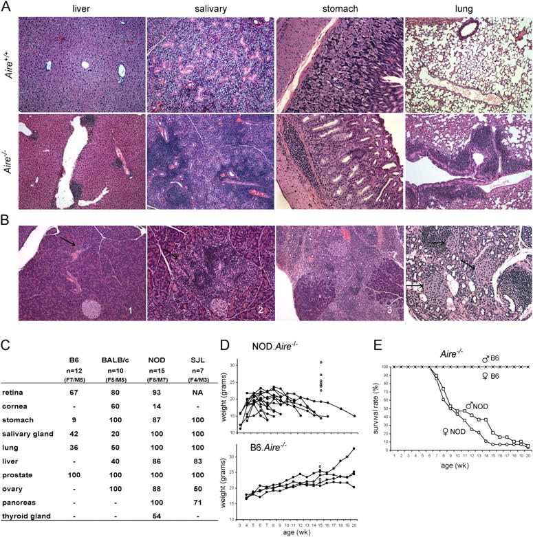
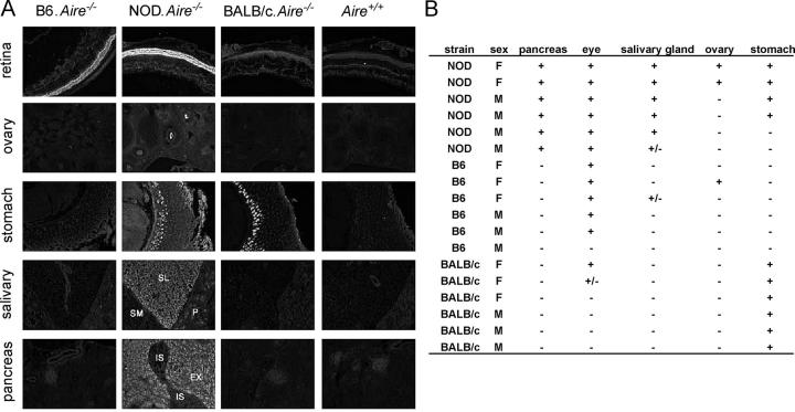
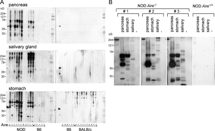
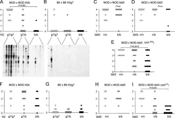
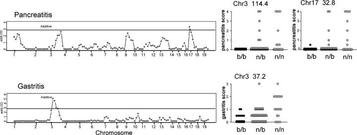
Loss of function mutations in the autoimmune regulator (Aire) gene in autoimmune polyendocrinopathy-candidiasis-ectodermal dystrophy patients and mutant mice lead to autoimmune manifestations that segregate as a monogenic trait, but with wide variation in the spectrum of organs targeted. To investigate the cause of this variability, the Aire knockout mutation was backcrossed to mice of diverse genetic backgrounds. The background loci strongly influenced the pattern of organs that were targeted (stomach, eye, pancreas, liver, ovary, thyroid, and salivary gland) and the severity of the targeting (particularly strong on the nonobese diabetic background, but very mild on the C57BL/6 background). Autoantibodies mimicked the disease pattern, with oligoclonal reactivity to a few antigens that varied between Aire-deficient strains. Congenic analysis and a whole genome scan showed that autoimmunity to each organ had a distinctive pattern of genetic control and identified several regions that controlled the pattern of targeting, including the major histocompatibility complex and regions of Chr1 and Chr3 previously identified in controlling type 1 diabetes.


Figures

Figure 1.
Figure 1.
The genetic background conditions the autoimmunity caused by Aire deficiency. (A) Lymphocytic infiltrates in Aire-deficient mice. Hematoxylin and eosin–stained liver (10× objective), salivary (10×), stomach (20×), and lung (10×) sections from 20-wk-old sex-matched littermates that were Aire+/+ (top) and Aire−/− (bottom) on an NOD (liver, salivary gland, and lung) or BALB/c background (stomach). (B) Pancreatic histology of NOD Aire−/− mice at various stages of disease. (1) Perivascular infiltration (arrow), (2) patches of infiltrates (arrow) in exocrine pancreas, (3) massive infiltration in exocrine pancreas, and (4) complete destruction of exocrine pancreas, whereas the islets are spared (black arrows). (C) Percent incidence of disease in different organs in Aire−/− mice on B6, BALB/c, NOD, and SJL backgrounds at 20 wk of age. Some of the NOD and SJL background mice were analyzed a few weeks earlier. n = total number of knockout mice analyzed. −, no affected mice. Retina refers to retinal degeneration. Except for NOD background mice, in which sialitis was detected in 2/5 Aire+/+ controls, Aire-proficient littermates showed rare and mild pathology (2/11 lung infiltrates on the B6, 1/5 salivary gland in SJL, and 1/5 ovary in BALB/c backgrounds, respectively). (D) Growth curves of NOD.Aire−/− and B6.Aire−/− females. Open circles at 15 wk are the weights of groups of Aire−/− littermates. (E) Survival of NOD.Aire+/+ females (open circles) and males (open squares; n = 28 and 19, respectively) and B6. Aire−/− mice (closed circles and squares; n = 7 and 7, respectively).
Figure 2.
Figure 2.
Autoantibodies from Aire-deficient mice of different genetic backgrounds match the disease specificity. (A) Representative staining of frozen sections from retina, ovary, stomach, salivary, and pancreas (10×) of a RAG-deficient mouse with sera from 10-wk-old Aire−/− mice from different backgrounds, and an age- and sex- matched Aire+/+ mouse (B6 background, except for stomach section [BALB/c background]). P, parotid; Sl, sublingual gland; Sm, submandibular gland; Ex, exocrine pancreas; Is, pancreatic islets. (B) Summary of the presence of organ-specific autoAbs to the indicated organs in individual 10-wk-old Aire−/−mice from different backgrounds. ±, trace staining.
Figure 3.
Figure 3.
Stronger and different autoAbs in NOD.Aire / than in other strains. (A) Each lane of the multiscreen immunoblot shows autoAbs in the serum from a 10- or 20-wk-old individual Aire −/− mouse (−) or WT littermate (+) on the NOD, B6, or BALB/c background immunoblotted with pancreatic, salivary, or gastric antigens. Anti–β-actin antibody as a positive control is in the first lane of each membrane. (B) Higher resolution immunoblot of autoAb targets in different tissues with sera from three different mice.
Figure 4.
Figure 4.
Different NOD congenic intervals influence Aire-associated pancreatitis and gastritis. NOD.Aire−/− and B6.Aire−/− mice were intercrossed with a set of congenic mice with different MHC or Idd intervals. (A, B, F, and G) MHC (reciprocal on B6 and NOD backgrounds). (C and H) Idd5 b on the NOD background. (D) Idd3 b on the NOD background. (E and I) Idd3 b on the NOD.Idd5 b background. Note that all NOD.Idd3 mice also carried the B6 allele at Idd5. The resulting Aire-deficient homozygous animals, which shared a genetic background except for the indicated genotype at the congenic interval, were scored histologically for pancreatitis (A–E) and gastritis (F–I) at 20 wk of age. Each circle represents an individual Aire−/− mouse. Statistical p-values were obtained with a Wilcoxon rank sum test. (A and B, bottom) Immunoblots from sera of animals of the indicated genotypes, probing pancreas extracts.
Figure 5.
Figure 5.
Pancreatitis and gastritis in intercrossed NOD.Aire / and B6.Aire / mice. (A) Pancreatitis and gastritis in Aire−/− (B6 × NOD) F1 mice. (B) Aire−/− (B6 × NOD) F2 mice were generated by intercrossing F1 parents, and the resulting mice were scored for disease. The biparametric plots show scores of individual mice for pancreatitis versus gastritis (left) and pancreatitis versus pneumonitis (right). (C) Antipancreas autoAbs were assayed by immunoblot in sera from (B6 × NOD) F2 Aire−/− mice. Each lane corresponds to an individual F2 mouse whose histological pancreatitis score is shown at the top.
Figure 6.
Figure 6.
Different genetic regions are associated with pancreatitis and gastritis in Aire / (B6 × NOD) F2 mice. 98 Aire−/− (B6 × NOD) F2 mice were tested histologically and genotyped across the entire genome (117 SNP markers distinguishing NOD and B6 genomes, displayed in chromosomal order). The LOD score is shown for association with pancreatitis (top) and gastritis (bottom). A score of 2.96 corresponds to a genome-wide significance of P = 0.05 (1,000 permutations). (right) Disease scores for animals grouped by genotype at the different markers are shown.

References

    1. Bach, J.F. 2002. The effect of infections on susceptibility to autoimmune and allergic diseases. N. Engl. J. Med. 347:911–920. - PubMed
    1. Kishimoto, H., and J. Sprent. 2001. A defect in central tolerance in NOD mice. Nat. Immunol. 2:1025–1031. - PubMed
    1. Liston, A., S. Lesage, D.H. Gray, L.A. O'Reilly, A. Strasser, A.M. Fahrer, R.L. Boyd, J. Wilson, A.G. Baxter, E.M. Gallo, et al. 2004. Generalized resistance to thymic deletion in the NOD mouse; a polygenic trait characterized by defective induction of Bim. Immunity. 21:817–830. - PubMed
    1. Choisy-Rossi, C.M., T.M. Holl, M.A. Pierce, H.D. Chapman, and D.V. Serreze. 2004. Enhanced pathogenicity of diabetogenic T cells escaping a non-MHC gene-controlled near death experience. J. Immunol. 173:3791–3800. - PubMed
    1. Zucchelli, S., P. Holler, T. Yamagata, M. Roy, C. Benoist, and D. Mathis. 2005. Defective central tolerance induction in NOD mice: genomics and genetics. Immunity. 22:385–396. - PubMed

Publication types

MeSH terms