. 2005 Sep 19;202(6):829-39.
doi: 10.1084/jem.20050199.

A role for TSLP in the development of inflammation in an asthma model

Affiliations

A role for TSLP in the development of inflammation in an asthma model

Amin Al-Shami et al. J Exp Med. .

Abstract

Thymic stromal lymphopoietin (TSLP) is a cytokine that promotes CD4+ T cell homeostasis. We now demonstrate that TSLP is required to mount a normal CD4+ T cell-mediated inflammatory response. TSLP acts directly on naive, but not, memory CD4+ T cells, and promotes their proliferation in response to antigen. In addition, TSLP exerts an effect indirectly through DCs to promote Th2 differentiation of CD4+ T cells. Correspondingly, TSLP receptor (TSLPR) knockout (KO) mice exhibit strong Th1 responses, with high levels of interleukin (IL)-12, interferon-gamma, and immunoglobulin (Ig) G2a, but low production of IL-4, -5, -10, -13, and IgE; moreover, CD4+ T cells from these animals proliferate less well in response to antigen. Furthermore, TSLPR KO mice fail to develop an inflammatory lung response to inhaled antigen unless supplemented with wild-type CD4+ T cells. This underscores an important role for this cytokine in the development of inflammatory and/or allergic responses in vivo.


Figures
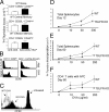
Figure 1.
Figure 1.
TSLP promotes the proliferation of naive CD4+ T cells. (A) Purified naive (CD4+CD62L+CD44), central memory (CD4+CD62L+CD44+), and effector memory (CD4+CD62LCD44+) T cells from WT BALB/c mice were activated for 48 h with 2 μg/ml anti-CD3 with or without 100 ng/ml TSLP and then pulsed with [3H]thymidine for 16 h. TSLP significantly increased the proliferation of naive CD4+ T cells (P = 0.01; top panel) but did not significantly affect memory CD4+ T cell proliferation (middle and bottom panels). (B) Defective CD69 induction in TSLPR KO mice. WT mice, and TSLPR KO mice expressing the DO11.10 transgene were injected i.p. with OVA and aluminum hydroxide (ALUM) and analyzed the next day. (C) Decreased antigen-induced CD4+ T cell proliferation in TSLPR KO mice. CD4+ T cells from DO11.10/WT and TSLPR KO mice were labeled with CFSE and transferred to WT hosts that were immunized with OVA the next day. On day 5, KJ1-26+ DO11.10/TSLPR KO cells had slower proliferation (mean fluorescence intensity (MFI) = 267 and percent gated 54%) than did DO11.10/WT cells (MFI = 12 and percent gated 98%) (cells from 7 out of 10 TSLPR KO mice showed a slower rate of CFSE loss than WT cells, whereas cells from 3 out of 10 did not). (D–F) WT and TSLPR KO mice (not expressing the DO11.10 transgene) were immunized with OVA (200 μg) and ALUM. Animals were killed on day 12 (D) or 60 (E) and splenocytes were cultured with OVA (0, 10, 50, and 200 μg/ml). Splenocytes from TSLPR KO mice displayed significantly lower proliferation in response to secondary encounter with antigen at all concentrations tested (P < 0.05 for all). (F) CD4+ T cells purified from mice 60 d after immunization were incubated with APCs (splenocytes that were depleted of T and NK cells) in the presence of different doses of OVA. CD4+ T cells from TSLPR KO mice displayed significantly lower proliferation in response to the OVA (200 μg/ml) in vitro (P < 0.01) than did CD4+ T cells from WT mice. Shown are means ± SEM for five experiments.
Figure 2.
Figure 2.
TSLP activates murine DCs. (A) In vitro antigen recall response. CD4+ T cells and APCs from immunized WT and TSLPR KO mice were cultured with 200 μg/ml of OVA for 48 h and then pulsed with [3H]thymidine. Shown are means ± SEM for seven experiments. (B) Splenic CD11c+ cells were sorted from the spleens of WT BALB/c animals and incubated overnight with media, TSLP alone, 5 μg/ml of OVA323–339 peptide, or with OVA323–339 peptide plus TSLP. TSLP treatment increased the surface levels of CD80, MHC class II, and CD86. Numbers represent mean fluorescent intensity. (C) Splenic DCs were incubated with 5 μg/ml of OVA323–339 peptide alone or with TSLP before being washed, treated with mitomycin C, and cultured with purified CD4+ T cells from DO11.10 RAG2−/− mice at a 1:10 ratio. Antigen presentation of DCs was enhanced significantly by TSLP treatment (P = 0.005, shown are means ± SEM for five experiments). (D) CD4+ T cells and DCs were purified from DO11.01/WT and DO11.10/TSLPR KO unmanipulated mice, and these were cultured together in the indicated combinations at a ratio of 1:10 DC/T cell, with 5 μg/ml of OVA323–339 peptide, and [3H]thymidine incorporation determined at 48 h. Replacing WT APCs with TSLPR KO-derived DCs moderately, but significantly, reduced the antigen-driven proliferation of DO11.10 Tg/WT CD4+ T cells (P = 0.04). WT DCs provided no significant enhancement over the weak expansion of DO11.10/TSLPR KO CD4+ T cells. Shown are means ± SEM for seven experiments. (A and D) Asterisks indicate that the difference between WT CD4+/WT APCs or DCs and WT CD4+/KO APCs or DCs was statistically significant (P < 0.05).
Figure 3.
Figure 3.
DCs activated with TSLP negatively regulate IFN-γ production by naive CD4+ T cells. In panels A, B, C, and E, cells were cultured as indicated and subsequently challenged with PMA+ ionomycin, and the intracellular levels of IFN-γ and IL-4 were measured by intracellular staining. Numbers indicate the percent of cells in the indicated quadrant. (A) Naive and memory CD4+ T cells (>99% pure) were isolated from WT animals and treated with anti-CD3 with or without TSLP for 4 d. The addition of TSLP had no effect on the levels of IFN-γ and IL-2 that were produced by CD4+ T cells. (B) Purified CD4+ T cells were activated with anti-CD3/anti-CD28 under Th1 (IL-12 and anti–IL-4) or Th2 (IL-4 and anti–IFN-γ) polarizing conditions with or without TSLP. IL-2 was added on day 2, and cells were allowed to grow for 1 wk. TSLP did not affect the IFN-γ or IL-4 production by these polarized cells. (C) Sorted splenic CD11c+ DCs were incubated with 5 μg/ml of OVA323–339 peptide alone or with TSLP before being washed, treated with mitomycin C, and cultured with purified CD4+ T cells from DO11.10 RAG2−/− mice at a 1:10 ratio from nonimmunized animals. TSLP-treated DCs reduced the levels of IFN-γ production by KJ1-26+CD4+ T cells, whereas the levels of IL-4 were not affected. (D) No significant difference was observed in the levels of IL-12 (p35) mRNA examined by RT-PCR in CD11c+ DCs that were incubated overnight with 5 μg/ml of OVA323–339 peptide alone or with TSLP. (E) Total splenocytes from DO11.10/WT and DO11.10/TSLPRKO mice were cultured for 4 d with 5 μg/ml of OVA323–339 peptide before being activated with PMA+ ionomycin for 5 h. KJ1-26+ TSLPR KO T cells produced more IFN-γ than did DO11.10/WT cells. (F) RNA was extracted from DO11.10/WT and DO11.10/TSLPRKO total splenocytes that were cultured for 4 d with 5 μg/ml of OVA323–339 peptide. RT-PCR revealed significantly lower levels of IL-4 mRNA in the spleens of TSLPR KO mice; asterisk indicates that the value was significantly lower (P < 0.05). The experiment was done four times with one or two mice in each experimental group in each experiment.
Figure 4.
Figure 4.
TSLPR KO mice fail to mount an inflammatory response. (A–D) Periodic acid-Schiff–stained lung tissue sections of BALB/c WT and TSLPR KO mice that were sensitized (i.p.) and challenged (i.t. and i.n.) with OVA or PBS (i.p.). There were no obvious differences in the lung morphology between WT (A) and TSLPR KO (C) animals that were exposed to PBS. WT mice that received OVA displayed perivascular inflammation, peribronchiolar cuffing, and goblet cell hyperplasia (B), whereas TSLPR KO mice that were treated with OVA showed no obvious inflammation (D). (E) WT and TSLPR KO animals treated as shown above were tested for their airway hypersensitivity response using methacholine (0, 6, 12, 25, and 50 mg/ml). Unlike WT animals, TSLPR KO mice immunized and sensitized with OVA showed similar results to PBS control mice (six to seven animals per group, shown is one representative experiment out of three). *Statistical significance (P < 0.05) between the indicated dose and the 0 mg/ml. (F) Splenocytes from immunized WT and TSLPR KO mice were cultured in vitro with 200 μg/ml OVA for 3 d. Supernatants were examined for IFN-γ and IL-4 levels. The levels of IL-4 were significantly lower in the TSLPR KO animals versus the WT animals, whereas no statistical significance was observed for IFN-γ. The experiment was done four times with four mice in each experimental group in each experiment. *Statistical significance (P < 0.05).
Figure 5.
Figure 5.
TSLPR KO mice exhibit partially altered allergic/ inflammatory immunoglobulin and cytokine profiles in the lungs in OVA-treated mice. BALB/c WT and TSLPR KO mice were sensitized (i.p.) and challenged (i.t. and i.n.) with OVA or PBS (i.p.). (A) Sera were tested at the indicated dilutions and the levels of OVA-specific immunoglobulin in PBS- and OVA-treated mice. Antibody was captured with plate-bound ovalbumin (2 μg/ml) and detected with biotinylated isotype-specific secondary antibodies. OVA-TSLPR KO mice displayed significantly less IgE and more IgG2a than did WT treated littermates. (B) Cytokine levels in the lungs as determined by RT-PCR. (C) Cytokines protein levels in BAL as determined by ELISA. The results are expressed as means ± SEM for four experiments, two to four mice per group per experiment. (A and B) *P < 0.05 between PBS and OVA group of WT and PBS and OVA group of TSLPR KO mice.
Figure 6.
Figure 6.
TSLP plays a role in the development of asthma. (A)TSLPR KO mice succeed in mounting an inflammatory response in the lung after receiving WT CD4+ T cells. Donor and recipient mice (F4 BALB/c) were sensitized with OVA (i.p.). Recipient mice were challenged with OVA (i.t.) 4 h before receiving 8 × 10⁶ CD4+ T cells. CD4+ T cells from WT donor were transferred to WT (positive control, i and ii) and TSLPR KO (test subject, panels iii and iv) mice, whereas CD4+ T cells from TSLPR KO mice were given to TSLPR KO mice to act as negative control (v and vi). All mice were treated the next day with OVA (i.n.) and killed 24 h later. Shown are the periodic acid-Schiff staining of lung tissue sections. The WT positive control displayed perivascular inflammation, peribronchiolar cuffing, and goblet cell hyperplasia (i and ii), whereas TSLPR KO mice showed no signs of lung inflammation (iii and iv). However, TSLPR KO mice that were supplemented with CD4+ T cells (v and vi) exhibited inflammatory cells infiltration combined with peribronchiolar cuffing, and goblet cell hyperplasia in the large and medium airways. (B) Lung sections stained with periodic acid-Schiff show the effect of blocking TSLP using Fc-TSLPR fusion protein on the lung inflammation in WT animals that were immunized and sensitized with OVA. Introducing Fc-TSLPR fusion protein 4 h before each of the i.t. and i.n. OVA reduced the levels of inflammation in these animals (v and vi, score 1.6 with a range of 1–2.5) compared with animals that were treated with Ig (iii and iv, score 2.6 and a range of 2–3.5). WT mice that were treated with OVA (i and ii) or PBS (vii and viii) acted as positive and negative controls, respectively. The difference was statistically significant (P = 0.01). The experiment was done three times with two mice in each experimental group in each experiment.

Similar articles

Cited by

References

    1. Friend, S.L., S. Hosier, A. Nelson, D. Foxworthe, D.E. Williams, and A. Farr. 1994. A thymic stromal cell line supports in vitro development of surface IgM+ B cells and produces a novel growth factor affecting B and T lineage cells. Exp. Hematol. 22:321–328. - PubMed
    1. Park, L.S., U. Martin, K. Garka, B. Gliniak, J.P. Di Santo, W. Muller, D.A. Largaespada, N.G. Copeland, N.A. Jenkins, A.G. Farr, et al. 2000. Cloning of the murine thymic stromal lymphopoietin (TSLP) receptor: formation of a functional heteromeric complex requires interleukin 7 receptor. J. Exp. Med. 192:659–670. - PMC - PubMed
    1. Pandey, A., K. Ozaki, H. Baumann, S.D. Levin, A. Puel, A.G. Farr, S.F. Ziegler, W.J. Leonard, and H.F. Lodish. 2000. Cloning of a receptor subunit required for signaling by thymic stromal lymphopoietin. Nat. Immunol. 1:59–64. - PubMed
    1. Leonard, W.J. 2001. Cytokines and immunodeficiency diseases. Nat. Rev. Immunol. 1:200–208. - PubMed
    1. Sims, J.E., D.E. Williams, P.J. Morrissey, K. Garka, D. Foxworthe, V. Price, S.L. Friend, A. Farr, M.A. Bedell, N.A. Jenkins, et al. 2000. Molecular cloning and biological characterization of a novel murine lymphoid growth factor. J. Exp. Med. 192:671–680. - PMC - PubMed

Publication types

MeSH terms