. 2005 Sep 27;102(39):14052-7.
doi: 10.1073/pnas.0506713102. Epub 2005 Sep 19.

The neuronal PAS domain protein 3 transcription factor controls FGF-mediated adult hippocampal neurogenesis in mice

Affiliations

The neuronal PAS domain protein 3 transcription factor controls FGF-mediated adult hippocampal neurogenesis in mice

Andrew A Pieper et al. Proc Natl Acad Sci U S A. .

Abstract

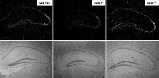
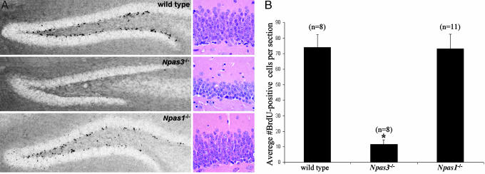
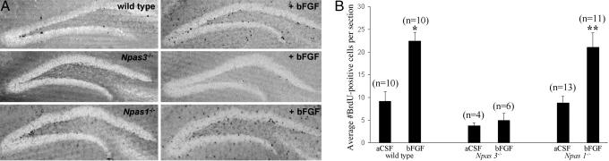
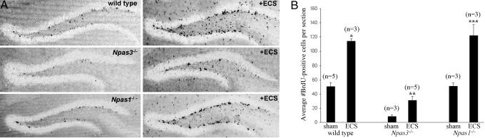
The neuronal PAS domain protein 3 (NPAS3) gene encoding a brain-enriched transcription factor was recently found to be disrupted in a family suffering from schizophrenia. Mice harboring compound disruptions in the NPAS3 and related NPAS1 genes manifest behavioral and neuroanatomical abnormalities reminiscent of schizophrenia. Herein we demonstrate that Npas3-/- mice are deficient in expression of hippocampal FGF receptor subtype 1 mRNA, most notably in the dentate gyrus. In vivo BrdUrd-labeling shows that basal neural precursor cell proliferation in the dentate gyrus of Npas3-/- mice is reduced by 84% relative to wild-type littermates. We propose that a deficiency in adult neurogenesis may cause the behavioral and neuroanatomical abnormalities seen in Npas3-/- mice, and we speculate that impaired neurogenesis may be involved in the pathophysiology of schizophrenia.


Figures

Fig. 1.
Fig. 1.
Dark-field illumination in situ hybridization pattern for FGF receptor subtype 1 (FGFR1) mRNA showing attenuation in the hippocampus of Npas3-/- mice relative to wild-type and Npas1-/- mice. Bright-field illumination shown directly below the in situ hybridization panels illustrates the neuroanatomy of each section
Fig. 2.
Fig. 2.
Basal neural precursor cell proliferation in the hippocampal dentate gyrus SGZ and GL showing statistically significant decreases in Npas3-/- (n = 8) adult mice relative to wild-type littermate (n = 8) and Npas1-/- (n = 11) animals (*, P < 0.0001, Student's t test). (A) Mice received daily i.p. injection of BrdUrd (50 μg/g of body weight) for 12 days and representative immunohistochemical staining of incorporated BrdUrd on day 13 is shown in hippocampal dentate gyrus of wild-type, Npas3-/-, and Npas1-/- adult mouse brain. Hematoxylin/eosin staining showed markedly reduced thickness of the dentate gyrus granular layer in Npas3-/- adult mice relative to wild-type and Npas1-/- adult mice. (B) Average numbers of BrdUrd-positive cells per section within the dentate gyrus were 74 ± 8 in wild-type mice, 12 ± 3 in Npas3-/- mice, and 73 ± 9 in Npas1-/- mice
Fig. 3.
Fig. 3.
Proliferation of neural precursor cells was increased in a statistically significant manner in the hippocampal dentate gyrus GL and SGZ of adult wild-type and Npas1-/- mice after lateral ventricle i.c.v infusion of bFGF dissolved in aCSF. Neural precursor cell proliferation in Npas3-/- mice was not affected by i.c.v. infusion of bFGF. (A) Representative immunohistochemical staining of BrdUrd is shown in hippocampal dentate gyrus of wild-type, Npas3-/-, and Npas1-/- adult mice after i.c.v. infusion of either aCSF vehicle control or bFGF. BrdUrd-positive cells were quantified within the dentate gyrus GL and SGZ in the hemisphere contralateral to the site of cannula placement to avoid false signal due to surgical artifact. (B) Average numbers of BrdUrd-positive cells per section significantly increased from 9 ± 2 in wild-type mice infused with aCSF alone (n = 10) to 22 ± 2 in wild-type mice infused with bFGF dissolved in aCSF (n = 10) (*, P < 0.0001, Student's t test), and from 9 ± 2 in Npas1-/- mice infused with aCSF alone (n = 13) to 21 ± 3 in Npas1-/- mice infused with bFGF dissolved in aCSF (n = 11) (**, P < 0.0001, Student's t test). Average numbers of BrdUrd-positive cells per section were 4 ± 1 in Npas3-/- mice infused with aCSF alone (n = 4) and 5 ± 2 in Npas3-/- mice infused with bFGF dissolved in aCSF (n = 6)
Fig. 4.
Fig. 4.
Proliferation of neural precursor cells was increased in a statistically significant manner in the hippocampal dentate gyrus of adult wild-type, Npas3-/-, and Npas1-/- mice after ECS. (A) Representative immunohistochemical staining of BrdUrd is shown in the hippocampal dentate gyrus of wild-type, Npas3-/-, and Npas1-/- mice after ECS. (B) Average numbers of BrdUrd-positive cells per section were increased in a statistically significant manner from 50 ± 6 in sham-ECS (n = 5) up to 121 ± 6 in ECS-treated (n = 3) wild-type mice (*, P < 0.001, Student's t test), from 9 ± 2 in sham-ECS (n = 3) up to 31 ± 6 in ECS-treated (n = 5) Npas3-/- mice (**, P < 0.001, Student's t test), and from 51 ± 5 in sham-ECS (n = 3) up to 122 ± 15 in ECS-treated (n = 3) Npas1-/- mice (***, P < 0.001, Student's t test)

Similar articles

Cited by

References

    1. Kamnasaran, D., Muir, W. J., Ferguson-Smith, M. A. & Cox, D. W. (2003) J. Med. Genet. 40, 325-332. - PMC - PubMed
    1. Brunskill, E. W., Witte, D. P., Shreiner, A. B. & Potter, S. S. (1999) Mech. Dev. 88, 237-241. - PubMed
    1. Zhou, Y.-D., Barnard, M., Tian, H., Li, X., Ring, H. Z., Franke, U., Shelton, J., Richardson, J., Russel, D. W. & McKnight, S. L. (1997) Proc. Natl. Acad. Sci. USA 94, 713-718. - PMC - PubMed
    1. Erbel-Sieler, C., Dudley, C., Zhou, Y., Wu, X., Estill, S. J., Han, T., Diaz-Arrastia, R., Brunskill, E. W., Potter, S. S. & McKnight, S. L. (2004) Proc. Natl. Acad. Sci. USA 101, 13648-13653. - PMC - PubMed
    1. Kempermann, G., Kuhn, H. G. & Gage, F. H. (1997) Nature 386, 493-495. - PubMed

Publication types

MeSH terms

Substances