. 2005 Sep 27;102(39):13909-14.
doi: 10.1073/pnas.0506517102. Epub 2005 Sep 19.

Breast cancer bone metastasis mediated by the Smad tumor suppressor pathway

Affiliations

Breast cancer bone metastasis mediated by the Smad tumor suppressor pathway

Yibin Kang et al. Proc Natl Acad Sci U S A. .

Erratum in

  • Proc Natl Acad Sci U S A. 2006 May 30;103(22):8570

Abstract

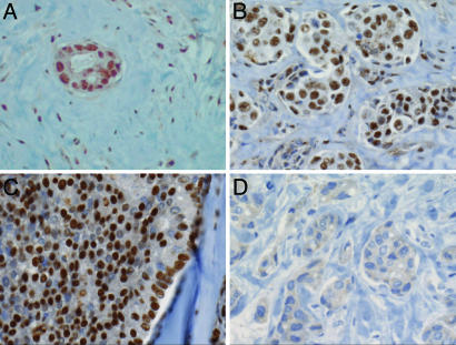
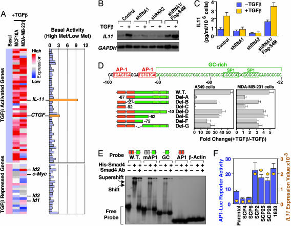
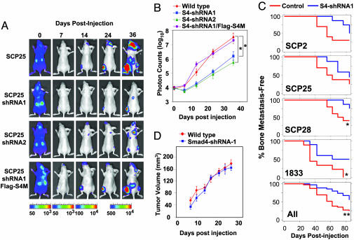
TGF-beta can signal by means of Smad transcription factors, which are quintessential tumor suppressors that inhibit cell proliferation, and by means of Smad-independent mechanisms, which have been implicated in tumor progression. Although Smad mutations disable this tumor-suppressive pathway in certain cancers, breast cancer cells frequently evade the cytostatic action of TGF-beta while retaining Smad function. Through immunohistochemical analysis of human breast cancer bone metastases and functional imaging of the Smad pathway in a mouse xenograft model, we provide evidence for active Smad signaling in human and mouse bone-metastatic lesions. Genetic depletion experiments further demonstrate that Smad4 contributes to the formation of osteolytic bone metastases and is essential for the induction of IL-11, a gene implicated in bone metastasis in this mouse model system. Activator protein-1 is a key participant in Smad-dependent transcriptional activation of IL-11 and its overexpression in bone-metastatic cells. Our findings provide functional evidence for a switch of the Smad pathway, from tumor-suppressor to prometastatic, in the development of breast cancer bone metastasis.


Figures

Fig. 1.
Fig. 1.
Activated Smad pathway in breast cancer bone metastasis. Examples of intense immunohistochemical staining of receptor-phosphorylated Smad2 in breast cancer bone metastasis samples from different patients. The samples shown were chosen to illustrate the nuclear phospho-Smad2 staining in a metastatic island and the surrounding stroma (A), in a cluster of metastatic islands (B), or in a contiguous metastatic mass (C), as well as a cluster of islands stained with normal rabbit serum as a negative control (D).
Fig. 2.
Fig. 2.
Functional imaging of Smad signaling in breast cancer bone metastasis. (A) Schematic representation of the retroviral vectors SFG-tdRFP-cmvFLuc (constitutively expressing tdRFP and FLuc) and cis-TGF-β1–Smads–HSV1-tk/GFP (expressing HSV-tk/GFP fusion protein in response to TGF-β). (B and C) SCP3 transduced with these two vectors were treated with TGF-β or no additions for 24 h and analyzed by fluorescence microscopy (B) or two-color FACS (C). The constitutive tdRFP fluorescence is shown on the ordinate, and the HSV-tk/GFP fusion fluorescence, inducible by TGF-β, is shown on the abscissa. (D and E Upper) In vivo bioluminescence and microPET imaging of metastases in mice. SCP2 (D) and SCP3 (E Upper) cells bearing the SFG-tdRFP-cmvFLuc and cis-TGF-β1–Smads–HSV1-tk/GFP vectors were injected into the left cardiac ventricle and analyzed after 4 weeks (SCP2) or 18 weeks (SCP3). Bioluminescence imaging shows sites of metastases in the skull (D and E) and adrenal gland (E Upper). 18F-2′-fluoro-2′deoxy-1β-D-arabionofuranosyl-5-ethyl-uracil microPET images of tk/GFP reporter activation shows localization of radioactivity to the skull in the coronal and sagittal image planes. No visualization of the adrenal metastasis was seen on microPET imaging. Note the nonspecific accumulation of the tracer in the gastrointestinal tract and bladder attributable to clearance of the tracer. (E Lower) At necroscopy, the head showing the skull and the adrenal metastasis plus kidney were removed and imaged ex vivo for photographic (–) and bioluminescence (+) imaging.
Fig. 3.
Fig. 3.
Smad4 and AP1-dependent transcriptional activation of IL-11 by TGF-β. (A) Basal expression levels of 50 TGF-β-activated genes and 21 TGF-β-repressed genes in MCF-10A and MDA-MB-231 cells were normalized to the same level. (Left) Responses of these genes to TGF-β in each cell line are represented by different shades of red (degrees of activation) or blue (degrees of repression) in the dendrogram. (Right) The ratio of basal expression levels of these 71 genes in highly metastatic versus weakly metastatic MDA-MB-231 cells. Genes of interest are highlighted. (B) SCP25 and its derivatives (see Fig. 5B) were incubated in the absence or presence of TGF-β for 2 h. Total RNA was subjected to Northern blot analysis with the indicated probes. (C) SCP25 and its derivatives were treated with or without TGF-β for 24 h. IL-11 production in the media was determined with an ELISA assay. Data are the average of triplicate determinations ± SD. (D)(Upper) Nucleotide sequence of the minimal TGF-β-responsive region of the IL-11 promoter. Nucleotide sequence positions are indicated relative to the transcription start site. Two AP1 sites (red boxes) and a GC-rich sequence (green) containing two SP1 sites (green boxes) are indicated. (Lower)(Center and Right) A549 (Center) and MDA-MB-231 (Right) cells were transfected with the indicated IL-11 reporter constructs, treated with or without TGF-β for 16–20 h before lysis, and analyzed for luciferase activlty. Data are the average of triplicate determinations ± SD. (Left) The schematic representation of each promoter construct. (E) [γ32-P]ATP end-labeled probes matching to the wild-type IL-11 proximal promoter region, this region with mutant AP1 sites (mAP1), or the indicated fragments of this region were subjected to electrophoretic mobility shift analysis with recombinant full-length His-Smad4 protein. Antibody against Smad4 was added as indicated to create supershifts. The β-actin promoter was used as a negative control. Schematic representations of the probes are shown above the gel. (F) Various MDA-MB-231 sublines were transfected with 1 μg of 4xAP1-Luc reporter plasmid and analyzed for luciferase activity 2 days after transfection. Data are the average of triplicate determinations ± SD. The absolute values of IL 11 mRNA level as detected by an Affymetrix U133A GeneChip were plotted in the same graph (yellow circles). The scales for the luciferase activity and for IL-11GeneChip expression values are shown to the left and right of the graph, respectively.
Fig. 4.
Fig. 4.
Smad4 mediation of breast cancer bone metastasis. Wild-type and genetically modified SCP25 was labeled with the TGL reporter and 1 × 10⁵ cells were injected into the left cardiac ventricle of five mice for each cell line. At the indicated days after xenografting, bioluminescence images were acquired and quantified. (A) Representative mice from each group are shown in the supine position. The intensity of the signal from days 24 and 36 are on equivalent scales, whereas days 0, 7, and 14 are each on separate scales because of increasing signal strength and to avoid signal saturation. (B) The normalized photon counts from the bone metastases in the hindlimbs were measured over the indicated time course. (C) Kaplan–Meier curves showing the incidence of bone metastasis by indicated wild-type and Smad4-knockdown MDA-MB-231 sublines. For each cell sample, 105 tumor cells were inoculated into the left cardiac ventricle of 10 nude mice. Metastasis was scored as the time to first appearance of a visible bone lesion by x-ray imaging of the whole mouse. The percentages of animals in each group and in all groups combined that were free of detectable bone metastases are plotted. *, < 0.05; **, P < 0.01; calculated by log rank test. (D) Tumor cells (106) were injected s.c. into nude mice. s.c. tumor growth was monitored and quantified by caliper measurements. No significant difference was found between wild-type and Smad4-knowdown cells.

Similar articles

Cited by

References

    1. Derynck, R., Akhurst, R. J. & Balmain, A. (2001) Nat. Genet. 29, 117–129. - PubMed
    1. Siegel, P. M. & Massague, J. (2003) Nat. Rev. Cancer 3, 807–821. - PubMed
    1. Shi, Y. & Massague, J. (2003) Cell 113, 685–700. - PubMed
    1. Watanabe, T., Wu, T. T., Catalano, P. J., Ueki, T., Satriano, R., Haller, D. G., Benson, A. B., III, & Hamilton, S. R. (2001) N. Engl. J. Med. 344, 1196–1206. - PMC - PubMed
    1. Siegel, P. M., Shu, W., Cardiff, R. D., Muller, W. J. & Massague, J. (2003) Proc. Natl. Acad. Sci. USA 100, 8430–8435. - PMC - PubMed

Publication types

MeSH terms

Substances