. 2005 Sep 27;102(39):13855-60.
doi: 10.1073/pnas.0504307102. Epub 2005 Sep 19.

NO-mediated cytoprotection: instant adaptation to oxidative stress in bacteria

Affiliations

NO-mediated cytoprotection: instant adaptation to oxidative stress in bacteria

Ivan Gusarov et al. Proc Natl Acad Sci U S A. .

Abstract

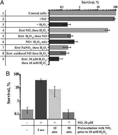
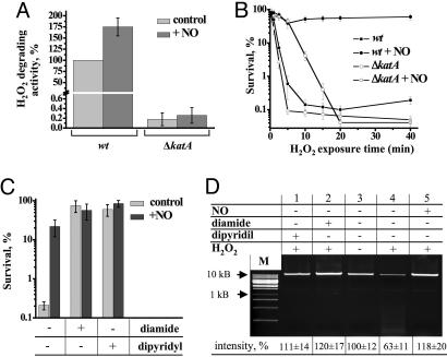
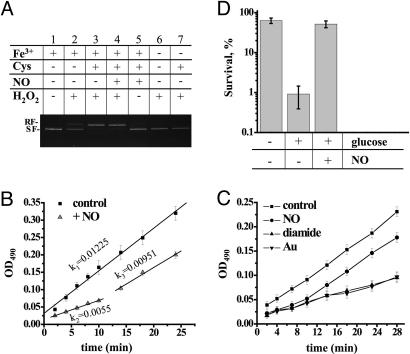
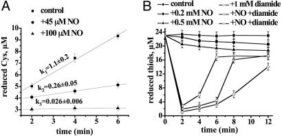
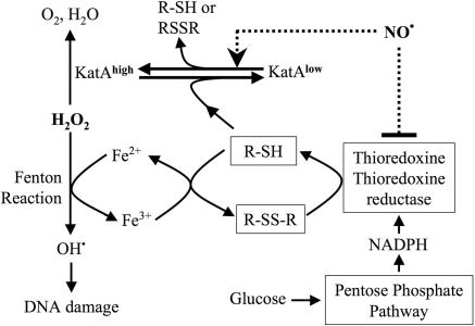
Numerous sophisticated systems have been described that protect bacteria from increased levels of reactive oxygen species. Although indispensable during prolonged oxidative stress, these response systems depend on newly synthesized proteins, and are hence both time and energy consuming. Here, we describe an "express" cytoprotective system in Bacillus subtilis which depends on nitric oxide (NO). We show that NO immediately protects bacterial cells from reactive oxygen species by two independent mechanisms. NO transiently suppresses the enzymatic reduction of free cysteine that fuels the damaging Fenton reaction. In addition, NO directly reactivates catalase, a major antioxidant enzyme that has been inhibited in vivo by endogenous cysteine. Our data also reveal a critical role for bacterial NO-synthase in adaptation to oxidative stress associated with fast metabolic changes, and suggest a possible role for NO in defending pathogens against immune oxidative attack.


Figures

Fig. 1.
Fig. 1.
Cytoprotection by exogenous NO. (A) Effect of exogenous NO on H2O2 toxicity. The final concentration of NO after a bolus application was 30 μM. The concentrations of H2O2 and NaNO2 were 10 mM and 30 μM, respectively. Reagents were added as indicated to aerobically grown wt cells at OD600 ∼ 0.5 (in LB at 37°C). Incubation with H2O2 was for 30 min. In lanes 4, 7, and 8, H2O2 was added 5 sec after NO, NaNO2, or oxidized NO, respectively. In lane 6, NO was mixed with H2O2 before addition to cells. In lane 8, NO was oxidized before addition to cells by bubbling air into an aqueous solution of NO for 2 h. In lane 9, 10 mM H2O2 was added after a 5-sec incubation with 30 μMH2O2. The percentage of surviving cells was determined by colony formation and is shown as the mean ± SD from five experiments. (B) Time course of NO-mediated cytoprotection. At the times shown after the addition of NO, aliquots of culture were removed and challenged with 10 mM H2O2 for 30 min. Cm (200 μg/ml) was added for 5 min before NO/H2O2 treatment. Values shown are the means ± SD from three experiments.
Fig. 2.
Fig. 2.
Activation of catalase and inhibition of the Fenton reaction, two components of NO-mediated cytoprotection. (A) Stimulating effect of NO on H2O2 degrading activity in crude extracts of wt and ΔkatA cells. Total H2O2 degrading activity was measured as described in Experimental Procedures (31). Where indicated, extracts were incubated with 45 μM NO for 5 sec. 100% catalase activity = 30 mM H2O2 min–1·mg–1. Values shown are the means ± SE from six experiments. (B) Transient protection of Δkat cells from oxidative stress by NO. The graph shows the time course of H2O2-mediated toxicity. Ten millimolar H2O2 was added at t = 0. Where indicated, 30 μM NO was added 5 sec before H2O2. Values shown are the means ± SD from three experiments. (C) Protection of wt cells from oxidative stress by the iron chelator dipyridyl and thiol oxidizer diamide. After 5 min of incubation with Cm (50 μg/ml), aerobically grown wt cells (OD600 ∼ 0.5) were treated with dipyridyl (1 mM for 10 min) or diamide (200 μM for 3 min) and/or NO (30 μM for 5 sec), followed by the addition of 10 mM H2O2 for 5 min. Values shown are the means ± SE from four experiments. (D) Chromosomal DNA damage from the Fenton reaction. A representative agarose gel shows a 10-kb PCR fragment amplified from B. subtilis chromosome. Chromosomal DNA was isolated from cells treated with diamide, dipyridyl, or NO and H2O2 as described in Figs. 1 A and 2C. M, 1-kb DNA ladder. The relative intensity of the full size DNA band is indicated at the bottom. Values shown are the means ± SD from three experiments.
Fig. 3.
Fig. 3.
Inhibition of Cys reduction is a mechanism of NO cytoprotection. (A) Effect of Cys and NO on Fenton-mediated DNA damage in vitro. As indicated, the supercoiled pBR322 plasmid (0.5 μg) was treated with 30 μM FeCl3, 10 mM Cys, 45 μM NO, or 10 mM H2O2 in 20 mM Tris·HCl buffer (pH 7.9). After a 10-min incubation at room temperature, the reaction was stopped and separated in a 1% agarose gel. RF, relaxed form; SF, supercoiled form. (B) NO-mediated suppression of cellular RE rereduction. The graph shows the negative effect of NO on the rate of formazan dye formation in the culture of wt cells grown in LB. Where indicated, cells were treated with 30 μM NO for 5 sec before the addition of 0.1 mg/ml MTS. Values shown are the means ± SE from three experiments. (C) Effects of NO, diamide, or TepAu on the rate of formazan dye accumulation. Conditions are as in B. TepAu and diamide were added 30 sec before MTS. Values shown are the means ± SD from three experiments. (D) Effect of carbon availability on oxidative stress survival and NO protection. Cells in mid-log phase were resuspended in M9 minimal medium containing 200 μg/ml Cm and incubated with or without glucose (50 mM for 15 min at 37°C). NO (30 μM) was added for 5 sec, followed by H2O2 (10 mM). Values shown are the means ± SD from three experiments.
Fig. 4.
Fig. 4.
Inhibition of Cys reduction by NO in vitro and in vivo.(A) Inhibition of Trx/TrxRed-mediated cystine reduction by NO in the reconstituted system (see Experimental Procedures). NO donor MAHMA (45 and 100 μM) was added before initiation of the reaction with NADPH. (B) NO transiently inhibits Cys reduction in vivo. Cells were treated with 1 mM diamide and/or the indicated amount of NO donor MAHMA. At the indicated time points, aliquots were withdrawn, and thiols were quantified as described in Experimental Procedures. NO added to the reaction after it was stopped did not affect the fluorescence yield (data not shown). Values shown are the means ± SD from three experiments.
Fig. 5.
Fig. 5.
Mechanism of NO-mediated protection from oxidative stress in B. subtilis. NO instantly protects cells from H2O2 toxicity by a dual mechanism. In the schematic shown, NO transiently interrupts the production of damaging hydroxyl radicals from the Fenton reaction by suppressing Cys reduction by Trx/TrxRed. In parallel, NO activates catalase (KatA).
Fig. 6.
Fig. 6.
NOS-mediated protection from oxidative stress in B. subtilis. (A) Effect of nos deletion on H2O2 sensitivity. wt and Δnos cells were grown aerobically in LB to late log phase (OD600 ∼ 0.8–0.9) at 30°C. An aliquot from each culture was diluted with an equal amount of fresh prewarmed LB for 2 min (diluted). Both diluted and undiluted aliquots were treated with 1 mM or 10 mM H2O2 for 30 min. The percentage of surviving cells was determined by colony formation. Values shown are the means and SD (error bars) from four independent experiments. (B) Exogenous Cys sensitizes B. subtilis to oxidative stress and induces NOS-mediated protection. Cells were grown as in A and diluted with an equal volume of saline or saline plus Cys (100 μM). Arg (100 μM) was added to all samples. After 2 min of incubation, cells were treated with 10 mM H2O2 for 30 min. Values shown are the means ± SD from three experiments. (C) Effect of fresh medium dilution on nitrite levels in wt and Δnos cell cultures. Conditions were as in A. Samples for nitrite measurements were taken 5 min after dilution. LB, nitrite level in LB. Values shown are the means ± SD from three experiments. (D) Effect of dilution on KatA activity. Conditions were as in A. Cells were collected 1 min after dilution and lysed immediately, and catalase activity was measured as described in Experimental Procedures. Values shown are the means ± SE from three experiments.

References

    1. Ignarro, L. J. (2002) J. Physiol. Pharmacol. 53, 503–514. - PubMed
    1. Moncada, S., Palmer, R. M. & Higgs, E. A. (1991) Pharmacol. Rev. 43, 109–142. - PubMed
    1. Kerwin, J. F., Jr., Lancaster, J. R., Jr., & Feldman, P. L. (1995) J. Med. Chem. 38, 4343–4362. - PubMed
    1. Nathan, C. & Shiloh, M. U. (2000) Proc. Natl. Acad. Sci. USA 97, 8841–8848. - PMC - PubMed
    1. Demple, B. (1999) Braz. J. Med. Biol. Res. 32, 1417–1427. - PubMed

Publication types

LinkOut - more resources