. 2005 Sep 27;102(39):13933-8.
doi: 10.1073/pnas.0501046102. Epub 2005 Sep 19.

An evolutionary proteomics approach identifies substrates of the cAMP-dependent protein kinase

Affiliations

An evolutionary proteomics approach identifies substrates of the cAMP-dependent protein kinase

Yelena V Budovskaya et al. Proc Natl Acad Sci U S A. .

Abstract

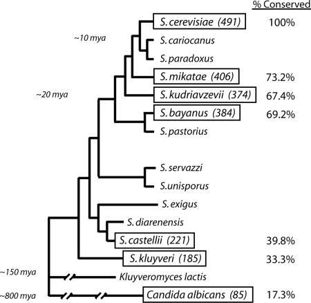
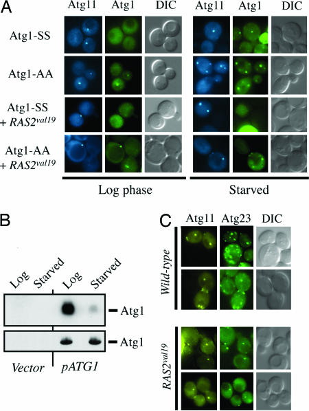
Protein kinases are important mediators of much of the signal transduction that occurs in eukaryotic cells. Unfortunately, the identification of protein kinase substrates has proven to be a difficult task, and we generally know few, if any, of the physiologically relevant targets of any particular kinase. Here, we describe a sequence-based approach that simplified this substrate identification process for the cAMP-dependent protein kinase (PKA) in Saccharomyces cerevisiae. In this method, the evolutionary conservation of all PKA consensus sites in the S. cerevisiae proteome was systematically assessed within a group of related yeasts. The basic premise was that a higher degree of conservation would identify those sites that are functional in vivo. This method identified 44 candidate PKA substrates, 5 of which had been described. A phosphorylation analysis showed that all of the identified candidates were phosphorylated by PKA and that the likelihood of phosphorylation was strongly correlated with the degree of target site conservation. Finally, as proof of principle, the activity of one particular target, Atg1, a key regulator of autophagy, was shown to be controlled by PKA phosphorylation in vivo. These data therefore suggest that this evolutionary proteomics approach identified a number of PKA substrates that had not been uncovered by other methods. Moreover, these data show how this approach could be generally used to identify the physiologically relevant occurrences of any protein motif identified in a eukaryotic proteome.


Figures

Fig. 1.
Fig. 1.
An evolutionary proteomics approach identified 85 potential substrates of PKA in S. cerevisiae. The relative number of S. cerevisiae candidates that have PKA sites conserved in the likely orthologous protein present in each of the indicated budding yeast species is shown. The approximate evolutionary distance to S. cerevisiae is shown for several positions on this phylogenetic tree (36, 49).
Fig. 2.
Fig. 2.
Proteins with highly conserved consensus PKA sites were more likely to be phosphorylated by PKA than proteins with less conserved sites. Shown is a phosphorylation analysis of representative proteins from the following five groups identified by the comparative analysis performed here: (i) proteins with consensus PKA sites conserved to C. albicans (A); (ii) proteins with sites conserved to the sensu lato and petite-negative Saccharomyces species; (iii) proteins with sites conserved amongst the sensu stricto Saccharomyces species (B); (iv) proteins with PKA sites in S. cerevisiae only (C); (v) proteins with no consensus PKA sites (D). For this analysis, the amount of label incorporated into a full-length GST fusion protein from [γ-32P]ATP was assessed with an in vitro assay performed in the presence or absence of PKA as described in Methods (26, 27). Note that the labeled protein bands have been appropriately lined up to facilitate comparisons between samples. Western immunoblots were performed with an α-GST antibody to quantify the relative amount of fusion protein present. (E) A summary graph indicating the percentage of candidates in each of the above groups that was phosphorylated by PKA.
Fig. 3.
Fig. 3.
The autophagy-related protein kinase, Atg1, is a substrate of PKA. (A) Elevated levels of Ras/PKA signaling activity inhibited autophagy. Autophagy levels were assessed with an alkaline phosphatase-based assay that has been described (24). The values shown represent the difference between the alkaline phosphatase levels found in starved and nonstarved cultures of the indicated yeast strains. HC-TPK1, high-copy plasmid encoding a catalytic subunit of PKA. (B) The full-length Atg1 was phosphorylated by PKA in vitro at the serine residues within the two consensus PKA sites. The residues at positions 508 and 515 within the two PKA sites are indicated: S, serine; A, alanine. Note that this experiment was performed with a kinase-inactive variant of Atg1, Atg1-K54A, to avoid the background autophosphorylation signal. (Lower) A Western immunoblot control indicating the relative levels of Atg1 present in each kinase reaction. (C and D) The in vitro (C) and in vivo (D) phosphorylation of an Atg1 fusion protein depended upon both PKA activity and the two PKA sites in Atg1. This fusion protein contained two repeats of the Ig-binding region of protein A fused in frame to residues 345–559 of Atg1 (28). In both cases, the Upper panel is the phosphorylation assay and the Lower is the Western immunoblot control. For the in vivo experiments, yeast cultures were incubated with [32P]orthophosphate, and the amount of label incorporated into the PrA-Atg1 fusion proteins was assessed by autoradiography. The pkaΔ strain lacks all three catalytic subunits of the S. cerevisiae PKA enzyme (33).
Fig. 4.
Fig. 4.
PKA phosphorylation regulates the association of Atg1 with the preautophagosomal structure, or PAS. (A) A fluorescence microscopy analysis of the subcellular localization of a YFP-Atg1 fusion protein in either log-phase or starved cultures of the indicated strains. The location of the PAS is shown by a CFP-Atg11 reporter construct (34). (B) The level of Atg1 phosphorylation decreased upon nitrogen starvation. Wild-type cells containing either a control vector (Vector) or a plasmid encoding a Protein A-Atg1 fusion protein (pATG1) were incubated with [32P]orthophosphate either before (Log) or after (Starved) a 2-h incubation in a medium lacking nitrogen. The amount of radiolabel incorporated into Atg1 was assessed by autoradiography. (Lower) The Western immunoblot control. (C) The presence of the RAS2val19 allele resulted in the redistribution of a GFP-Atg23 fusion protein from a number of punctate structures within the cell to the PAS. The identity of these punctate structures is not yet known. The location of the PAS is indicated by an RFP-Atg11 fusion protein.

Similar articles

Cited by

References

    1. Cohen, P. (2002) Nat. Rev. Drug Discov. 1, 309–315. - PubMed
    1. Hunter, T. (2000) Cell 100, 113–127. - PubMed
    1. Hunter, T. (1987) Cell 50, 823–829. - PubMed
    1. Manning, G., Whyte, D. B., Martinez, R., Hunter, T. & Sudarsanam, S. (2002) Science 298, 1912–1934. - PubMed
    1. Manning, B. D. & Cantley, L. C. (2002) Sci. STKE 162, 1–4. - PubMed

Publication types

MeSH terms

Substances