. 2005 Sep 27;102(39):13992-7.
doi: 10.1073/pnas.0504979102. Epub 2005 Sep 19.

Pathogenic myelin oligodendrocyte glycoprotein antibodies recognize glycosylated epitopes and perturb oligodendrocyte physiology

Affiliations

Pathogenic myelin oligodendrocyte glycoprotein antibodies recognize glycosylated epitopes and perturb oligodendrocyte physiology

Cecilia B Marta et al. Proc Natl Acad Sci U S A. .

Abstract

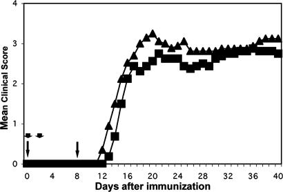
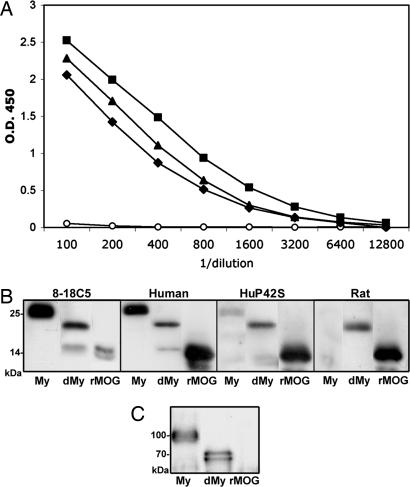
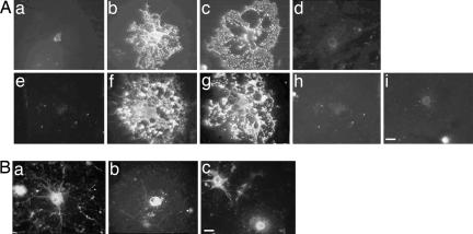
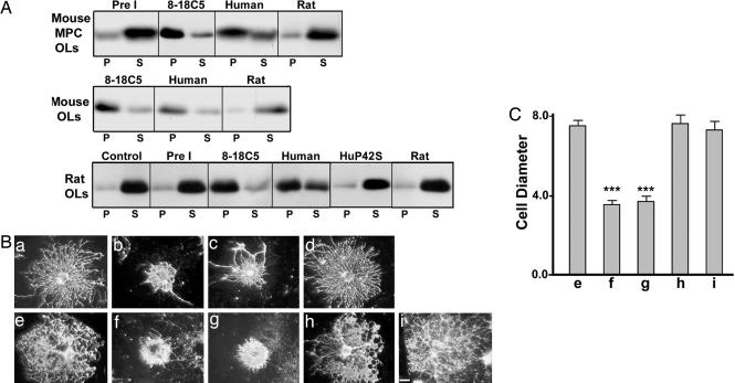
Antibodies to myelin components are routinely detected in multiple sclerosis patients. However, their presence in some control subjects has made it difficult to determine their contribution to disease pathogenesis. Immunization of C57BL/6 mice with either rat or human myelin oligodendrocyte glycoprotein (MOG) leads to experimental autoimmune encephalomyelitis (EAE) and comparable titers of anti-MOG antibodies as detected by ELISA. However, only immunization with human (but not rat) MOG results in a B cell-dependent EAE. In this study, we demonstrate that these pathogenic and nonpathogenic anti-MOG antibodies have a consistent array of differences in their recognition of antigenic determinants and biological effects. Specifically, substituting proline at position 42 with serine in human MOG (as in rat MOG) eliminates the B cell requirement for EAE. All MOG proteins analyzed induced high titers of anti-MOG (tested by ELISA), but only antisera from mice immunized with unmodified human MOG were encephalitogenic in primed B cell-deficient mice. Nonpathogenic IgGs bound recombinant mouse MOG and deglycosylated MOG in myelin (tested by Western blot), but only pathogenic IgGs bound glycosylated MOG. Only purified IgG to human MOG bound to live rodent oligodendrocytes in culture and, after cross-linking, induced repartitioning of MOG into lipid rafts, followed by dramatic changes in cell morphology. The data provide a strong link between in vivo and in vitro observations regarding demyelinating disease, further indicate a biochemical mechanism for anti-MOG-induced demyelination, and suggest in vitro tools for determining autoimmune antibody pathogenicity in multiple sclerosis patients.


Figures

Fig. 1.
Fig. 1.
Substitution of a proline with serine in human MOG overcomes the requirement for B cells to induce EAE. Wild-type (WT; rectangles) or μMT (triangles) mice were immunized as indicated in Methods and evaluated for clinical signs for 40 days. All mice in both groups developed EAE with a mean day of onset of 14.6 (WT) and 13 (μMT). Mortality was 0 of 8 (WT) and 2 of 10 (μMT). Mean maximum score was 3.2 (2.5–3.5) for WT and 3.7 (2.5–5) for μMT. Day 40 disease index (sum of mean clinical scores/day of onset × 100) was 463 for WT and 593 for μMT. Arrows, 100-μg MOG plus complete Freund's adjuvant; arrowheads, 500-ng pertussis toxin
Fig. 2.
Fig. 2.
IgGs from mice immunized with human, rat, or huP42S MOG, though equivalent by ELISA, differentially bind glycosylated MOG from purified myelin. (A) Purified IgG from pooled sera from mice 14 days after immunization with human (rectangles) or rat (diamonds) MOG protein, or 21 days after immunization with huP42S MOG protein (triangles), but not IgG from unimimmunized mice (circles), bound recombinant mouse MOG with approximately equivalent titer (ELISA; samples adjusted to same protein concentration). (B) Immunoblot of mouse myelin (My), deglycosylated myelin (dMy), or recombinant mouse MOG (rMOG): encephalitogenic mAb 8-18C5 and IgG from mice immunized with human, but not rat or huP42S, MOG protein bind MOG in purified myelin. In contrast, all four IgGs bind deglycosylated MOG in myelin and recombinant mouse MOG. (C) Immunoblot with anti-myelin associated glycoprotein (MAG): upon deglycosylation, ≈100-kDa MAG separates into bands of 72 (L-MAG) and 67 (S-MAG) kDa, respectively. Results are representative of three independent experiments
Fig. 3.
Fig. 3.
IgGs from mice immunized with mAb 8-18C5 or human, but not rat or huP42S, MOG protein bind to live OLs. (A) Mouse (ad) and rat (ei) OL cultures were incubated with preimmune IgG (a and e), 8-18C5 (b and f), or IgG purified from mice immunized with human (c and g), rat (d and h), or huP42S (i) MOG protein. (Bar, 5 μm.) (B) IgGs from mice immunized with human (a), rat (b), or huP42S (c) MOG protein bind intracellular MOG in fixed/permeabilized OLs. (Bar, 5 μm.) Results are representative of three independent experiments
Fig. 4.
Fig. 4.
MOG cross-linking with IgGs from mice immunized with human, but not rat or huP42S, MOG protein induces MOG repartitioning into a detergent insoluble fraction and morphological alterations in OLs. (A) MOG immunoblot of detergent soluble (S) or insoluble pellet (P) fractions from mixed primary or purified OL cultures incubated with media alone (Control), IgG from naïve mice (Pre I), mAb 8-18C5, or IgG from mice immunized with either human, huP42S, or rat MOG protein, followed by cross-linking with secondary antibodies. Treatment with mAb 8-18C5 or IgG raised against human MOG, but not huP42S or rat, MOG protein induces repartitioning of MOG into the detergent insoluble fraction. (B) Mouse (ad) or rat (ei) OLs were incubated with preimmune IgG (a and e), 8-18C5 (b and f), or IgG from mice immunized with human (c and g), rat (d and h), or huP42S (i) MOG protein, followed by cross-linking with anti-mouse IgG and stained with mAb O4 to visualize OL morphology. (Bar, 5 μm.) (C) Diameter (arbitrary units; mean ± SEM) of randomly chosen cells. IgG raised against human, but not rat or huP42S, MOG protein induces retraction of OL processes similar to 8-18C5 (***, P < 0.0001). Results are representative of three independent experiments

Similar articles

Cited by

References

    1. Haase, C. G., Guggenmos, J., Brehm, U., Andersson, M., Olsson, T., Reindl, M., Schneidewind, J. M., Zettl, U. K., Heidenreich, F., Berger, T., et al. (2001) J. Neuroimmunol. 114, 220-225. - PubMed
    1. Lindert, R. B., Haase, C. G., Brehm, U., Linington, C., Wekerle, H. & Hohlfeld, R. (1999) Brain 122, 2089-2100. - PubMed
    1. Genain, C. P., Cannella, B., Hauser, S. L. & Raine, C. S. (1999) Nat. Med. 5, 170-175. - PubMed
    1. Lassmann, H. (2004) in Myelin Biology and Disorders, ed. Lazzarini, R. A. (Elsevier Academic, San Diego), Vol. 2, pp. 733-762.
    1. Lucchinetti, C., Bruck, W., Parisi, J., Scheithauer, B., Rodriguez, M. & Lassmann, H. (2000) Ann. Neurol. 47, 707-717. - PubMed

Publication types

MeSH terms

LinkOut - more resources