. 2005 Sep 27;102(39):14075-9.
doi: 10.1073/pnas.0505340102. Epub 2005 Sep 19.

Rapid inactivation of a moth pheromone

Affiliations

Rapid inactivation of a moth pheromone

Yuko Ishida et al. Proc Natl Acad Sci U S A. .

Abstract

We have isolated, cloned, and expressed a male antennae-specific pheromone-degrading enzyme (PDE) [Antheraea polyphemus PDE (ApolPDE), formerly known as Sensillar Esterase] from the wild silkmoth, A. polyphemus, which seems essential for the rapid inactivation of pheromone during flight. The onset of enzymatic activity was detected at day 13 of the pupal stage with a peak at day 2 adult stage. De novo sequencing of ApolPDE, isolated from day 2 male antennae by multiple chromatographic steps, led to cDNA cloning. Purified recombinant ApolPDE, expressed by baculovirus, migrated with the same mobility as the native protein on both native polyacrylamide and isoelectric focusing gel electrophoresis. Concentration of ApolPDE (0.5 microM) in the sensillar lymph is approximately 20,000 lower than that of a pheromone-binding protein. Native and recombinant ApolPDE showed comparable kinetic parameters, with turnover number similar to that of carboxypeptidase and substrate specificity slightly lower than that of acetylcholinesterase. The rapid inactivation of pheromone, even faster than previously estimated, is kinetically compatible with the temporal resolution required for sustained odorant-mediated flight in moths.


Figures

Fig. 1.
Fig. 1.
Esterase activity in tissues of the wild silkmoth as visualized by α-/β-naphthyl acetate staining. ApolPDE (arrow) appears specifically in male antennae. MA, male antenna; I, integument; F, fat body; M, muscle; MG, midgut; MT, Malpighian tubules; HG, hindgut; C, central nervous system; T, testis; H, hemolymph
Fig. 2.
Fig. 2.
Development of male antennae and expression of ApolPDE during pupal-adult stages of the wild silkmoth. (A) Male antennae from pupae at 0, 2, 4, 6, 8, 10, 12, 13, and 14 days after incubation at room temperature and at day 0 adult. (Bar, 2 mm.) (B) To compare expression of ApolPDE, proteins were extracted from both male and female antennae at the same developmental stages as in A, separated by gel electrophoresis, and visualized by enzymatic activity
Fig. 3.
Fig. 3.
Purification of ApolPDE by ion exchange and gel filtration chromatography. (A and B) Anion exchange chromatography (DEAE and MonoQ, respectively). (C) Gel filtration (Superdex 75). (D) MonoQ (more shallow gradient). The horizontal bars at the bottom of each chromatogram in B–D indicate the collected fractions. (Insets) Proteins from indicated fractions were separated by 10% native PAGE and stained with α-/β-naphthyl acetate (A–C) or Coomassie blue (D). Arrowhead and arrows indicate the migration of ApolPDE
Fig. 4.
Fig. 4.
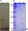
Separation of native ApolPDE by isoelectric focusing. (A) Crude antennal extract (lane 1) and a small aliquot (≈1 ng) of the purified native ApolPDE (lane 2) were separated by isoelectric focusing and visualized by enzymatic activity. (B) pI standards (lane 1) and the remainder of the isolated protein were separated on other lanes of the same gel and stained with Coomassie blue. Lane 2 was left empty to avoid contamination by the standard. Arrow indicates the isolated enzyme
Fig. 5.
Fig. 5.
Gene expression detected by RT-PCR. (A) Expression in male antennae and lack of expression of ApolPDE in female antennae and control tissues. I, integument; F, fat body; M, muscle; MG, midgut; MT, Malpighian tubules; HG, hindgut; T, testis; MA, male antenna; and FA, female antenna. (B) ApolPDE gene expression in male antennae started at day 13 of the pupal stage, 1 day earlier than the onset of another olfactory gene, ApolPBP1. Expression of ApolPBP1 has been demonstrated to start before adult eclosion (30). Previously identified genes (ApolODE, ApolIE) started expression early in the pupal stage. Actin, internal control

References

    1. Vogt, R. G. & Riddiford, L. M. (1981) Nature 293, 161-163. - PubMed
    1. Clyne, P. J., Warr, C. G., Freeman, M. R., Lessing, D., Kim, J. & Carlson, J. R. (1999) Neuron 22, 327-338. - PubMed
    1. Vosshall, L. B., Amrein, H., Morozov, P. S., Rzhetsky, A. & Axel, R. (1999) Cell 96, 725-736. - PubMed
    1. Sakurai, T., Nakagawa, T., Mitsuno, H., Mori, H., Endo, Y., Tanoue, S., Yasukochi, Y., Touhara, K. & Nishioka, T. (2004) Proc. Natl. Acad. Sci. USA 101, 16653-16658. - PMC - PubMed
    1. Leal, W. S., Chen, A. M., Ishida, Y., Chiang, V. P., Erickson, M. I., Morgan, T. I. & Tsuruda, J. M. (2005) Proc. Natl. Acad. Sci. USA 102, 5386-5391. - PMC - PubMed

Publication types

Associated data

LinkOut - more resources