The start site of the Acanthamoeba castellanii ribosomal RNA transcription unit
- PMID: 1617304
- PMCID: PMC6057357
The start site of the Acanthamoeba castellanii ribosomal RNA transcription unit
Abstract
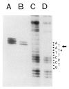
The 39S ribosomal RNA (rRNA) precursor has been isolated from Acanthamoeba castellanii. In vitro capping of the isolated RNA verified that it is the primary transcript and identified the 5' nucleotide as pppA. The position of the 5' coding nucleotide on the rRNA repeat unit sequence was identified using Northern blot, R-loop, and S1 nuclease mapping techniques. Dinucleotide priming of an in vitro transcription system stalled because of low initiating nucleotide concentration revealed that ApA maximally stimulates initiation of transcription. All of these results show that the underlined A in the sequence 5'-TATATATAAAGGGAC (RNA-like strand) coincides with the 5' nucleotide of the primary transcript. This identification is compatible with in vitro transcription experiments mapping the promoter for this transcription unit. The initiation sequences of rRNA genes from 14 species are compared, and a weak consensus for the initiator derived: [Formula; see text].
Figures


Similar articles
-
Sequence organization of the Acanthamoeba rRNA intergenic spacer: identification of transcriptional enhancers.Nucleic Acids Res. 1994 Nov 11;22(22):4798-805. doi: 10.1093/nar/22.22.4798. Nucleic Acids Res. 1994. PMID: 7984432 Free PMC article.
-
The ribosomal RNA gene region in Acanthamoeba castellanii mitochondrial DNA. A case of evolutionary transfer of introns between mitochondria and plastids?J Mol Biol. 1994 Jun 17;239(4):476-99. doi: 10.1006/jmbi.1994.1390. J Mol Biol. 1994. PMID: 8006963
-
The mitochondrial DNA of the amoeboid protozoon, Acanthamoeba castellanii: complete sequence, gene content and genome organization.J Mol Biol. 1995 Feb 3;245(5):522-37. doi: 10.1006/jmbi.1994.0043. J Mol Biol. 1995. PMID: 7844823
-
Regulation of ribosomal RNA transcription during differentiation of Acanthamoeba castellanii: a review.J Protozool. 1983 May;30(2):211-4. doi: 10.1111/j.1550-7408.1983.tb02905.x. J Protozool. 1983. PMID: 6355452 Review.
-
Initiation and regulation mechanisms of ribosomal RNA transcription in the eukaryote Acanthamoeba castellanii.Mol Cell Biochem. 1991 May 29-Jun 12;104(1-2):119-26. doi: 10.1007/BF00229811. Mol Cell Biochem. 1991. PMID: 1921990 Review.
Cited by
-
Sequence organization of the Acanthamoeba rRNA intergenic spacer: identification of transcriptional enhancers.Nucleic Acids Res. 1994 Nov 11;22(22):4798-805. doi: 10.1093/nar/22.22.4798. Nucleic Acids Res. 1994. PMID: 7984432 Free PMC article.
-
Two internal sequence elements modulate transcription from the external human 7S K RNA gene promoter in vivo.Gene Expr. 1999;8(2):105-14. Gene Expr. 1999. PMID: 10551798 Free PMC article.
-
Survey and summary: transcription by RNA polymerases I and III.Nucleic Acids Res. 2000 Mar 15;28(6):1283-98. doi: 10.1093/nar/28.6.1283. Nucleic Acids Res. 2000. PMID: 10684922 Free PMC article. Review.
-
TATA box-binding protein (TBP) is a constituent of the polymerase I-specific transcription initiation factor TIF-IB (SL1) bound to the rRNA promoter and shows differential sensitivity to TBP-directed reagents in polymerase I, II, and III transcription factors.Mol Cell Biol. 1994 Jan;14(1):597-605. doi: 10.1128/mcb.14.1.597-605.1994. Mol Cell Biol. 1994. PMID: 8264628 Free PMC article.
-
Promoter opening (melting) and transcription initiation by RNA polymerase I requires neither nucleotide beta,gamma hydrolysis nor protein phosphorylation.Nucleic Acids Res. 1993 Jul 11;21(14):3233-8. doi: 10.1093/nar/21.14.3233. Nucleic Acids Res. 1993. PMID: 7688114 Free PMC article.
References
Publication types
MeSH terms
Substances
Grants and funding
LinkOut - more resources
Full Text Sources
