. 2005 Sep 28;127(38):13213-9.
doi: 10.1021/ja051784p.

RNA catalysis in model protocell vesicles

Affiliations

RNA catalysis in model protocell vesicles

Irene A Chen et al. J Am Chem Soc. .

Abstract

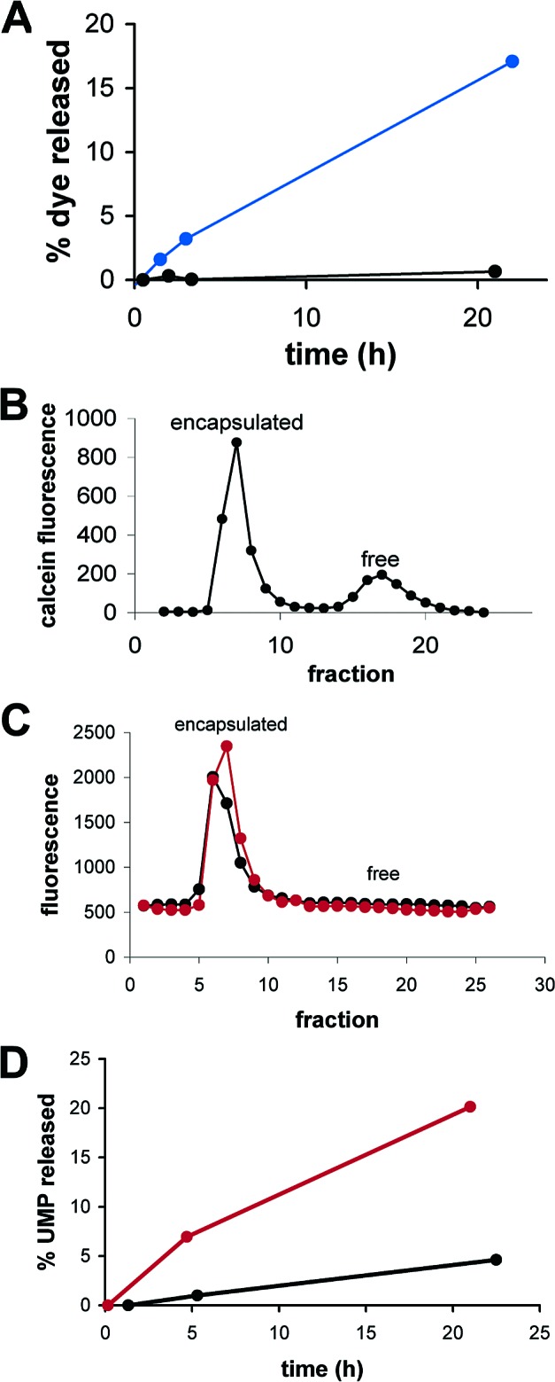
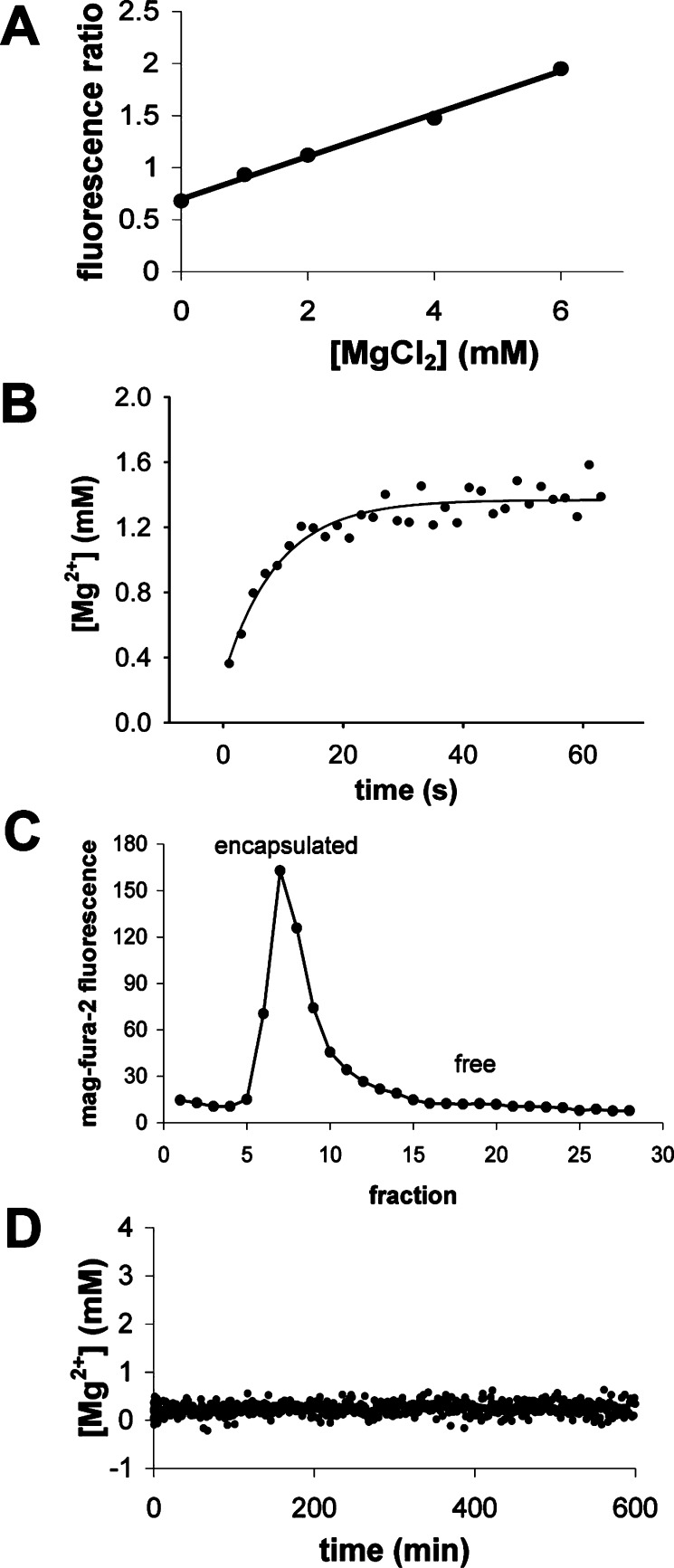
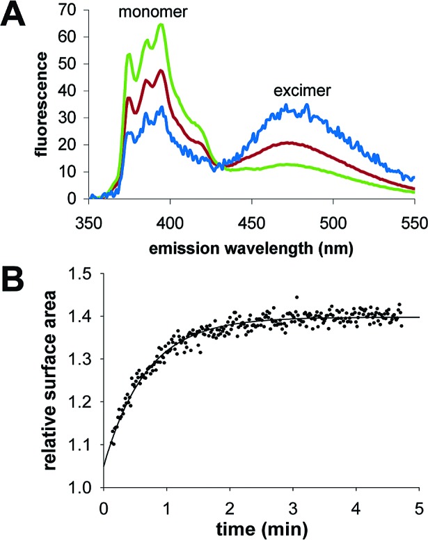
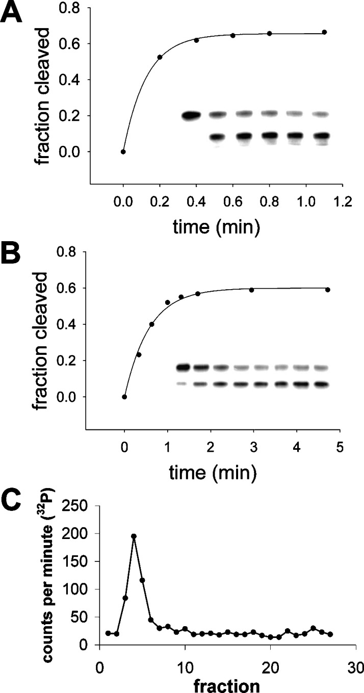
We are engaged in a long-term effort to synthesize chemical systems capable of Darwinian evolution, based on the encapsulation of self-replicating nucleic acids in self-replicating membrane vesicles. Here, we address the issue of the compatibility of these two replicating systems. Fatty acids form vesicles that are able to grow and divide, but vesicles composed solely of fatty acids are incompatible with the folding and activity of most ribozymes, because low concentrations of divalent cations (e.g., Mg(2+)) cause fatty acids to precipitate. Furthermore, vesicles that grow and divide must be permeable to the cations and substrates required for internal metabolism. We used a mixture of myristoleic acid and its glycerol monoester to construct vesicles that were Mg(2+)-tolerant and found that Mg(2+) cations can permeate the membrane and equilibrate within a few minutes. In vesicles encapsulating a hammerhead ribozyme, the addition of external Mg(2+) led to the activation and self-cleavage of the ribozyme molecules. Vesicles composed of these amphiphiles grew spontaneously through osmotically driven competition between vesicles, and further modification of the membrane composition allowed growth following mixed micelle addition. Our results show that membranes made from simple amphiphiles can form vesicles that are stable enough to retain encapsulated RNAs in the presence of divalent cations, yet dynamic enough to grow spontaneously and allow the passage of Mg(2+) and mononucleotides without specific macromolecular transporters. This combination of stability and dynamics is critical for building model protocells in the laboratory and may have been important for early cellular evolution.
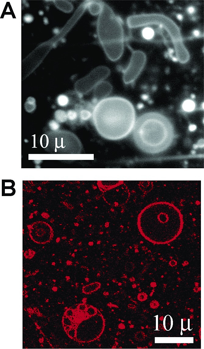

Figures

Figure 1
Figure 1
Leakage from MA:GMM vesicles (2:1) in 0.2 M bicine, pH 8.5, 4 mM MgCl2. (A) Vesicles were initially purified away from unencapsulated calcein, and dye leakage was measured over time with (blue) or without (black) the addition of 4 mM MgCl2. (B) Calcein leakage assayed by size-exclusion chromatography after 22 h in the presence of 4 mM MgCl2. (C) RNA leakage assayed by size-exclusion chromatography after 19 h, with (red) or without (black) the addition of 4 mM MgCl2. (D) Vesicles encapsulating 0.1 mM 5‘-UMP and a trace amount of 3H-UMP were initially purified, and then leakage of 3H-UMP was measured over time, with (red) or without (black) the addition of 4 mM MgCl2.
Figure 2
Figure 2
Mg2+ permeability in MA:GMM vesicles (2:1). (A) Standard curve of mag-fura-2 fluorescence ratio as a function of [Mg2+] in 0.2 M bicine, pH 8.5. The solid line is a linear regression fit. MgCl2 was added to a final concentration of 2 mM, by stopped-flow mixing, to MA:GMM (2:1) vesicles (B) or POPC vesicles (D) encapsulating mag-fura-2. The internal concentration of Mg2+ at each time point was calculated from the standard curve. The solid line (B) indicates a curve fit to a single-exponential equation, where k = 0.11 s-1. (C) Size-exclusion chromatography shows that no mag-fura-2 dye has leaked out of the MA:GMM vesicles after equilibration of Mg2+.
Figure 3
Figure 3
Growth of mixed composition vesicles by micelle incorporation. (A) Pyrene fluorescence shifts from monomer to excimer if aggregates are present. Shown are the spectra of pyrene in pure MA micelles (green), pure MA vesicles (blue), and MA:GMM (4:1) micelles (red). (B) Growth of 2:1:0.3 MA:GMM:dodecane vesicles over time, after addition of 1 equiv of micelles, in 0.2 M bicine, pH 8.5, 1 mM MgCl2. Relative surface area was determined using the FRET assay. The solid line indicates a curve fit to a single-exponential equation, with k = 1.5 min-1. A similar yield was obtained if 1 equiv of micelles was added at once or in up to five separate aliquots.
Figure 4
Figure 4
Self-cleavage activity of the hammerhead ribozyme N15min7. The insets show phosphorimages of the assay gels. The top band corresponds to the uncleaved ribozyme; the bottom band is the cleavage product. (A) Unencapsulated ribozyme activity in 0.2 M bicine, pH 8.5, 4 mM MgCl2. Time points, from left to right:  0 (no MgCl2), 0.2, 0.4, 0.6, 0.8, 1.1 min. The solid line indicates a curve fit to a single-exponential equation, y = a(1 − e-kx), where the extent of cleavage a = 0.66, and the observed rate constant k = 7.9 min-1. (B) Activity of ribozymes encapsulated in 2:1:0.3 MA:GMM:dodecane vesicles (∼15 mM amphiphile), in 0.2 M bicine, pH 8.5, 4 mM MgCl2. Time points, from left to right:  0 (no MgCl2), 0.33, 0.63, 1, 1.3, 1.7, 3, 4.7 min. Curve fit:  a = 0.6, and k = 1.7 min-1. (C) Size-exclusion chromatography of MA:GMM:dodecane vesicles shows that all radiolabeled N15min7 RNA remained encapsulated 15 min after the addition of MgCl2.
Figure 5
Figure 5
Fluorescence microscopy of 2:1:0.3 MA:GMM:dodecane vesicles containing hammerhead ribozyme in the presence of 3 mM MgCl2, prior to extrusion. Membranes were stained using Rhodamine 6G for visualization by (A) epifluorescence or (B) laser scanning confocal microscopy. The images shown are from different fields of view.

Similar articles

Cited by

References

    1. Szathmary E.; Demeter L. J. Theor. Biol. 1987, 128, 463–486. 10.1016/S0022-5193(87)80191-1. - DOI - PubMed
    1. Hogeweg P.; Takeuchi N. Origins Life Evol. Biosphere 2003, 33, 375–403. 10.1023/A:1025754907141. - DOI - PubMed
    1. Szabo P.; Scheuring I.; Czaran T.; Szathmary E. Nature 2002, 420, 340–343. 10.1038/nature01187. - DOI - PubMed
    1. Wade M. J. Proc. Natl. Acad. Sci. U.S.A. 1976, 73, 4604–4607. 10.1073/pnas.73.12.4604. - DOI - PMC - PubMed
    1. Wilson D. S. Proc. Natl. Acad. Sci. U.S.A. 1975, 72, 143–146. 10.1073/pnas.72.1.143. - DOI - PMC - PubMed

Publication types