. 2005 Sep-Oct;7(5):440-57.
doi: 10.1111/j.1525-142X.2005.05048.x.

Morphoregulation of teeth: modulating the number, size, shape and differentiation by tuning Bmp activity

Affiliations

Morphoregulation of teeth: modulating the number, size, shape and differentiation by tuning Bmp activity

Maksim V Plikus et al. Evol Dev. 2005 Sep-Oct.

Abstract

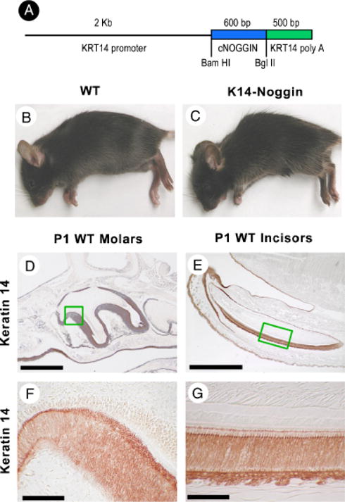
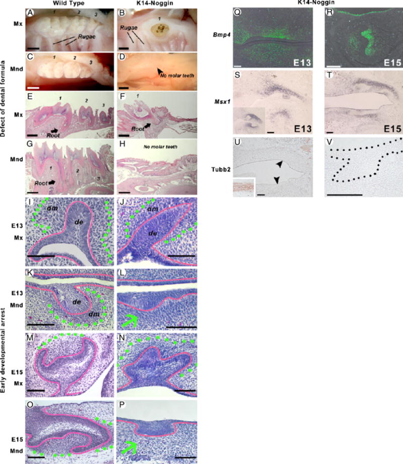
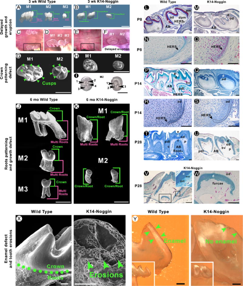
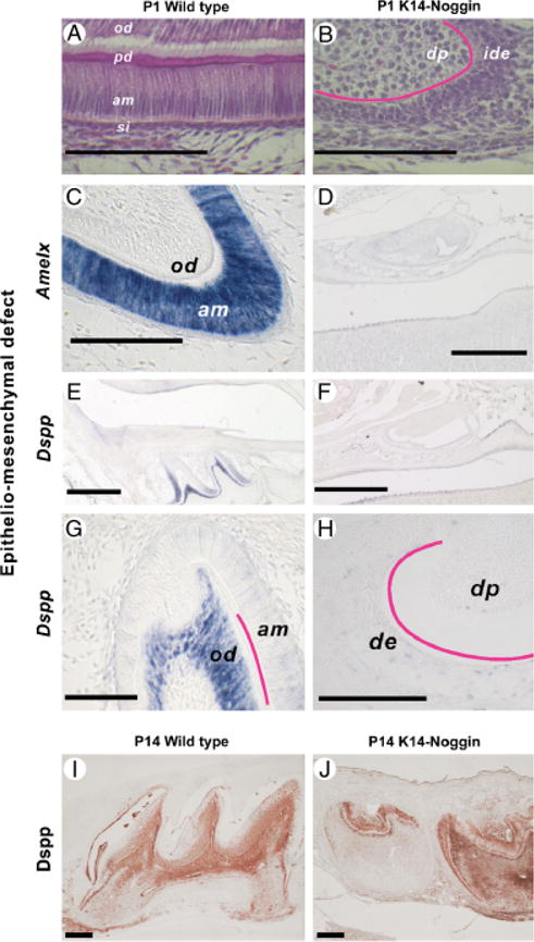
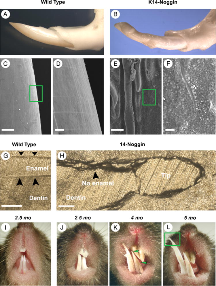
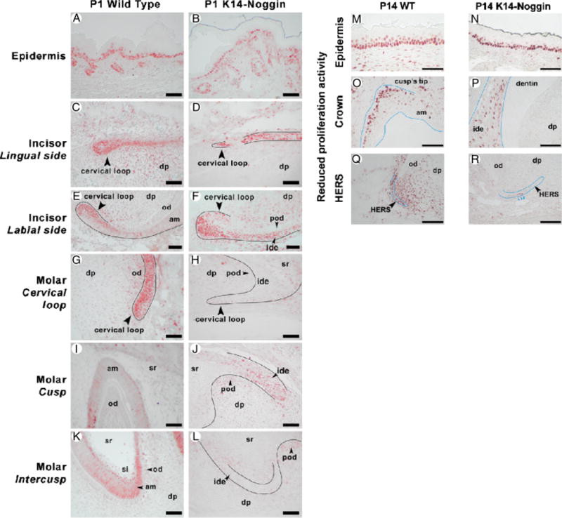
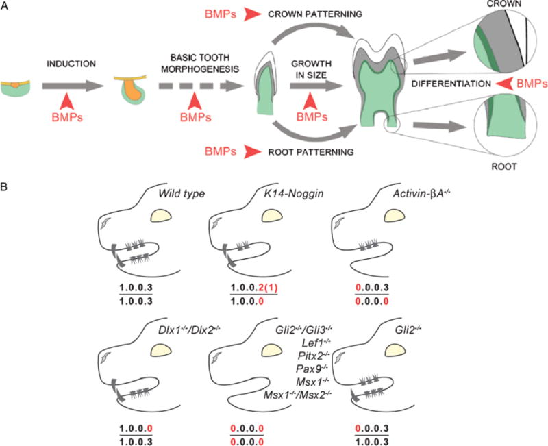
During development and evolution, the morphology of ectodermal organs can be modulated so that an organism can adapt to different environments. We have proposed that morphoregulation can be achieved by simply tilting the balance of molecular activity. We test the principles by analyzing the effects of partial downregulation of Bmp signaling in oral and dental epithelia of the keratin 14-Noggin transgenic mouse. We observed a wide spectrum of tooth phenotypes. The dental formula changed from 1.0.0.3/1.0.0.3 to 1.0.0.2(1)/1.0.0.0. All mandibular and M3 maxillary molars were selectively lost because of the developmental block at the early bud stage. First and second maxillary molars were reduced in size, exhibited altered crown patterns, and failed to form multiple roots. In these mice, incisors were not transformed into molars. Histogenesis and differentiation of ameloblasts and odontoblasts in molars and incisors were abnormal. Lack of enamel caused misocclusion of incisors, leading to deformation and enlargement in size. Therefore, subtle differences in the level, distribution, and timing of signaling molecules can have major morphoregulatory consequences. Modulation of Bmp signaling exemplifies morphoregulation hypothesis: simple alteration of key signaling pathways can be used to transform a prototypical conical-shaped tooth into one with complex morphology. The involvement of related pathways and the implication of morphoregulation in tooth evolution are discussed.


Figures

Fig. 1
Fig. 1
Production of K14-Noggin mouse. K14 Noggin construct used to generate transgenic mouse. The size of inset used and restriction enzyme are indicated (A). Appearance of control C57BL/6J (B) and mutant K14-Noggin 2.5-month-old mice (C). Examples of K14 immunostaining is shown in newborn molars (D, F) and incisors (E, G) which are strongly K14 positive. Section plane: sagittal (A–H). Scale bars: 100 μm (E); 50 μm (D); 10 μm (F, G).
Fig. 2
Fig. 2
Early arrest of K14-Noggin molar development. (A–D) WT mice have three maxillary (A) and three mandibular molars (C). Most of the K14-Noggin mice have only two or rarely one maxillary molar (B). None of the K14-Noggin mice have mandibular molars (D). (E–H) WT mice have three maxillary (E) and three mandibular molars (G) with a well-developed crown and roots. K14-Noggin mice have a reduced number of maxillary molars (F) and no mandibular molars. Instead, there is thickened oral epithelium, often resembling residual molar lamina (H). (I–L) Developing molars in the E13 WT (I, K) and K14-Noggin (J, L) mouse embryos. In WT mice, tooth buds have subjacent mesenchymal condensations (I, K). In K14-Noggin mice, the maxillary molar bud has a distinct mesenchymal condensation (outlined with green dotted line). However, the mandibular molar bud (L) lacks a mesenchymal condensation. Epithelium is outlined with red. (M–P) E15 molars in the WT (M, O) and K14-Noggin (N, P) mouse embryo. Unlike maxillary molars (N), K14-Noggin mandibular molars fail to develop further (P). Epithelium resembles initial tooth lamina (red). There is no mesenchymal condensation beneath it (green arrow). (Q, R) Bmp4 expression in the mesenchyme of the developing K14-Noggin teeth at E13 (Q) and E15 (R). (S, T) Msx1 expression pattern during early odontogenesis in K14-Noggin mice. In WT mice at E13, Msx1 is distinctly expressed in the dental mesenchyme, but not in the epithelium (S, inset). In E13 K14-Noggin mice, Msx1 is expressed in the dental mesenchyme of the maxillary molars (S). Msx1 expression is seen in the mandible, but not directly underneath the oral epithelium (S). At E15, when K14-Noggin mandibular molars show developmental arrest, Msx1 is expressed in the maxillary dental mesenchyme, as well as in the mesenchyme of the arrested mandibular early bud (T). (U, V) Absence of the neuro-specific differentiation of the dental mesenchyme in both maxillary and developmentally arrested mandibular tooth buds (V), as judged by the Tubb3 expression. Fibers of the trigeminal nerve are strongly positive for Tubb3 (see inset on U). Section plane: sagittal (E–H, J, L, N, P, Q–V), frontal (I, K M, O). Scale bars: 1 mm (A–H), 100 μm (I–V). de, dental epithelium; dm, dental mesenchyme.
Fig. 3
Fig. 3
Growth, eruption, and patterning and differentiation defects in K14-Noggin molars. (A, B) Comparative morphology of the maxillary molars in the P21 WT (A) and K14-Noggin mice (B). WT molars have a well-developed crown. M1 and M2 WT molars have well-developed multiple roots. Unlike WT teeth, maxillary M1 and M2 molars in K14-Noggin mice are smaller, have no clear crown to root separation, and do not form multiple roots. Postnatal development of the K14-Noggin maxillary molars is largely retarded. (C–F) Delayed eruption of molars in K14-Noggin mice. Although P21 M1 and M2 WT molars have fully erupted (C, D), both maxillary molars in P21 K14-Noggin mice remain at the early stages of eruption, with the crown mostly seated deep within the alveolae (E, F). (G–I) Abnormal crown pattering in K14-Noggin maxillary molars. P21 WT maxillary molars have a well-defined cusp pattern, with seven cusps in M1 and five cusps in the M2 molar (G). P21 K14-Noggin M1 and M2 maxillary molars have a severely abnormal crown pattern with small cusp-like prominences (H). Position and number of the intercusps are inconsistent in P21 M2 K14-Noggin molars (inverted view, I). (J, K) K14-Noggin molars have multiple root defects. WT molars have multiple well-developed roots (J). K14-Noggin molars have either two short, misconfigured roots (M1), or only one, very short root (M2), and multiple, irregular, grape-like growths on their surface (K). In WT molars, root length dominates over crown length (J). In K14-Noggin molars, the crown/root to multiple root ratio is changed and the crown/root length dominates over multiple root length (K). (L–S) Comparative morphology of P8 (L–O), P14 (P–S) WT, and K14-Noggin maxillary molars. Both in WT and K14-Noggin molars HERS is clearly seen at P8 (N, O). At P14 HERS in K14-Noggin molars has largely normal morphology (S vs. R). Although roots start to form at or before P8, furcae does not form until much later in K14-Noggin molars. (T–W) Formation of small furcae in P28 K14-Noggin molars. In M1 K14-Noggin molars furcae is first seen at P28 (V). Furcae is delineated by a layer of disorganized, mineralized dentin (W). Periodontal apparatus forms with periodontal ligament connected to the cementum (V, inset). Not all K14-Noggin molars form furcae and multiple roots (U vs. T). (X, Y) Crown surface defect in K14-Noggin molars. Unlike in WT molars, the crown surface of the K14-Noggin adult molars is uneven and highly eroded (X). On ground sections, WT molars show a distinct layer of enamel (Y, green arrows), whereas no enamel is seen in K14-Noggin molars (Y). Section plane: sagittal (L–W, Y). Scale bars: 1 mm (A, B, G, H, J, K); 0.5 mm (I); 200 μm (L, M, P, Q, T, U); 100 μm (V, X, Y), 50 μm (N, O, R, S, W). AB, aleveolar bone; am, ameloblasts; bv, blood vessels; c, cementum; d, dentin; dpm, dental papillae mesenchyme; e, enamel; f, furcae; HERS, Hertwig’s epithelial root sheath; od, odontoblasts; p, pulp; pdl, periodontal ligament.
Fig. 4
Fig. 4
Epithelio-mesenchymal defect in K14-Noggin molars. (A– D) Crown surface defect in K14-Noggin molars. Unlike in WT molars (A), the crown surface of the K14-Noggin adult molars is uneven and highly eroded (B). On ground sections, WT molars show a distinct layer of enamel (C, green arrows), whereas no enamel is seen in K14-Noggin molars (D). (A, B) Epithelium– mesenchyme interface of the P1 WT (A) and K14-Noggin (B) M1 molars. Unlike WT, K14-Noggin molars stay largely undifferentiated. (C–J) Absence of ameloblast-specific and delayed expression of odontoblast-specific markers. In P1 WT teeth, Amelx is strongly expressed in the ameloblasts, especially in the cusp (C). Dspp has strong expression in the preodontoblasts and odontoblasts, as well as in the preameloblasts of the intercusp (E, G). In P1 K14-Noggin molars, both Amelx (D) and Dspp (F, H) are largely absent. K14-Noggin molars gain Dspp expression later. Strong Dspp expression is seen at P14 (I vs. J). Scale bars: 500 μm (D–F); 200 μm (I, J); 100 μm (A–C, G, H). Section plane: sagittal (A–J). am, ameloblasts; de, dental epithelium; dp, dental papilla; ide, inner dental epithelium; od, odontoblasts; pd, predentine; si, stratum intermedium.
Fig. 5
Fig. 5
Growth abnormalities of the K14-Noggin incisors caused by the loss of enamel. (A, B) Comparative gross morphology of the mandibular incisors in the adult WT (A) and K14-Noggin mice (B). K14-Noggin incisors are thick, wide, blunt ended, and misaligned. (C–F) On SEM, the surface of the K14-Noggin incisors is rough and defective (E, F). It shows both macroscopic signs of deterioration in the form of deep, parallel ridges (E) and microscopic irregularities in the form of multiple bud-like formations (F). The surface of WT incisors is smooth (C, D). (G, H) On ground sections, WT incisors display a clear, thick layer of enamel (G). In contrast, K14-Noggin incisors do not have any enamel layer present (H). (I–L) Progressive changes of the incisors in K14-Noggin mice. Unlike WT incisors (I), K14-Noggin incisors are a dull white and deteriorate because of constant rubbing against each other (J–L). These changes start early in life and with age become more severe. The bottom incisors grew very long and became needle sharp (L). Section plane: sagittal (G, H). Scale bars: 100 μm (C, E, G, H); 20 μm (D, F).
Fig. 6
Fig. 6
Differentiation defect in K14-Noggin incisors. (A, B) Epithelium–mesenchyme interface of the P1 WT (A) and K14-Noggin (B) M1 incisors from the labial side. Unlike WT incisors, K14-Noggin incisors remain less differentiated. They do not form a distinct layer of polarized ameloblasts and there is no dentin or enamel deposition. Epithelium–mesenchyme interface is uneven and wavy. (C–J) Absence of the tooth-specific differentiation markers in the early postnatal (P1) K14-Noggin incisors. Amelx is strongly expressed in the ameloblasts of the WT incisors (C, D). In WT P1 molars Dspp has distinct expression (G). Dspp is expressed in the preodontoblasts and odontoblasts, as well as in the preameloblasts of the proximal labial side (H). In K14-Noggin P1 incisors, both Amelx (E, F) and Dspp (I, J) are largely absent. (K, L) Epithelium–mesenchyme interface of the P14 WT (K) and K14-Noggin (L) M1 incisors from the labial side. K14-Noggin incisors remain poorly differentiated. Epithelium–mesenchyme interface is uneven and wavy. Section plane: sagittal (A–L). Scale bars: 100 μm (A–L). am, ameloblasts; dp, dermal papilla; od, odontoblasts; pam, preameloblasts; pod, preodontoblasts; pd, predentine; si, stratum intermedium.
Fig. 7
Fig. 7
Proliferation defect in K14-Noggin teeth. (A, B) Similar rates of proliferation in the epidermis and hair follicles of P1 WT (A) and K14-Noggin (B) mice. (C–F) Proliferation pattern in P1 WT (C, E) and K14-Noggin (D, F) incisors. Proliferation rates are comparable on the lingual side of the cervical loop of both WT (C) and K14-Noggin (D) incisors. However, the proliferation zone on the labial side of the K14-Noggin cervical loop is significantly expanded distally (F vs. E). (G–L) Proliferation pattern in P1 WT (G, I, K) and K14-Noggin (H, J, L) molars. K14-Noggin molars show greatly reduced rates of proliferation, especially in the cervical loop (H vs. G) and intercusp area (L vs. K). (M–R) Reduced and nonlocalized proliferation activity in P14 K14-Noggin molars. WT molars show a localized zone of proliferation at the tip of the cusps (O) and extensive proliferation activity within HERS and the surrounding dental mesenchyme (Q). In contrast, K14-Noggin molars have reduced, de-centralized proliferation activity within the dental epithelium of the crown (P), largely reduced proliferation in HERS, and virtually no proliferation in the surrounding dental mesenchyme (R). Contrarily, the oral epithelium, adjacent to teeth, shows comparable proliferation activity both in WT (M) and K14-Noggin mice (N). Section plane: sagittal (A–R). Scale bars: 50 μm (A–R). am, ameloblasts; dp, dental papilla; ide, inner dental epithelium; od, odontoblasts; pod, preodontoblasts; si, stratum intermedium, sr, stellate reticulum.
Fig. 8
Fig. 8
Summary of the multiple dental defects caused by the disruption of the Bmp pathway in the oral epithelium. (A) Summary diagram of the tooth developmental events affected by the reduced strength of Bmp signaling in K14-Noggin mice. (B) Comparison of the dental formulas between WT, K14-Noggin, and other known mutant mice with reduction in teeth number.

Similar articles

Cited by

References

    1. Aberg T, Wozney J, Thesleff I. Expression patterns of bone morphogenetic proteins (bmps) in the developing mouse tooth suggest poles in morphogenesis and cell differentiation. Dev Dyn. 1997;210:383–396. - PubMed
    1. Andl T, et al. Epithelial Bmpr1a regulates differentiation and proliferation in postnatal hair follicles and is essential for tooth development. Development. 2004;131:2257–2268. - PubMed
    1. Begue-Kirn C, Krebsbach PH, Bartlett JD, Butler WT. Dentin sialoprotein, dentin phosphoprotein, enamelysin and ameloblastin: tooth-specific molecules that are distinctively expressed during murine dental differentiation. Eur J Oral Sci. 1998;106:963–970. - PubMed
    1. Bei M, Kratochwil K, Maas RL. BMP4 rescues a non-cell-autonomous function of Msx1 in tooth development. Development. 2000;127:4711–4718. - PubMed
    1. Bei M, Maas R. FGFs and BMP4 induce both Msx1-independent and Msx1-dependent signaling pathways in early tooth development. Development. 1998;125:4325–4333. - PubMed

Publication types

LinkOut - more resources