. 2005 Sep 20:2:21.
doi: 10.1186/1742-2094-2-21.

5-aminoimidazole-4-carboxamide-1-beta-4-ribofuranoside (AICAR) attenuates the expression of LPS- and Abeta peptide-induced inflammatory mediators in astroglia

Affiliations

5-aminoimidazole-4-carboxamide-1-beta-4-ribofuranoside (AICAR) attenuates the expression of LPS- and Abeta peptide-induced inflammatory mediators in astroglia

Kamesh R Ayasolla et al. J Neuroinflammation. .

Abstract

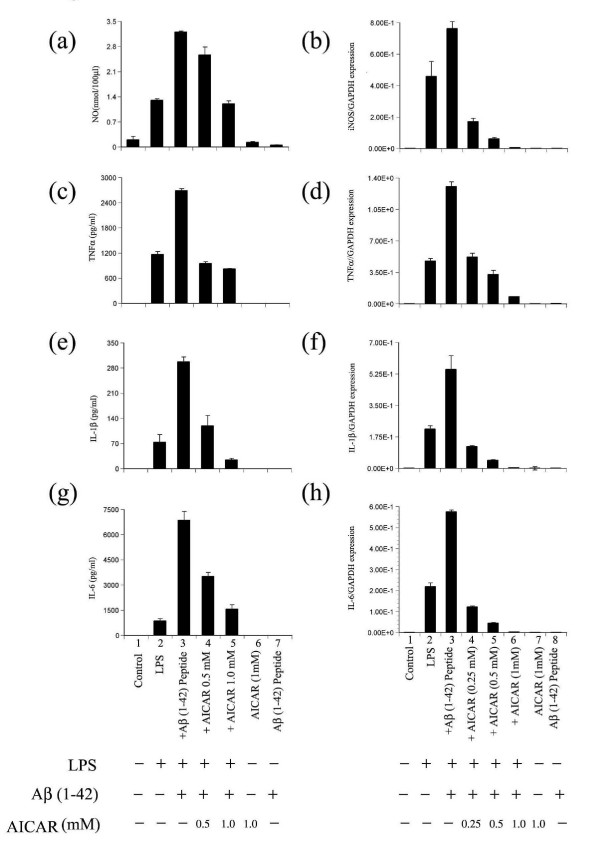
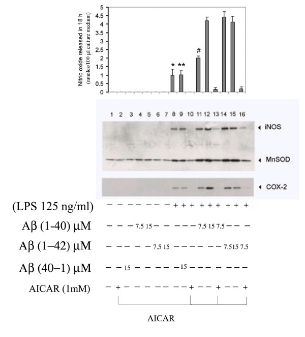
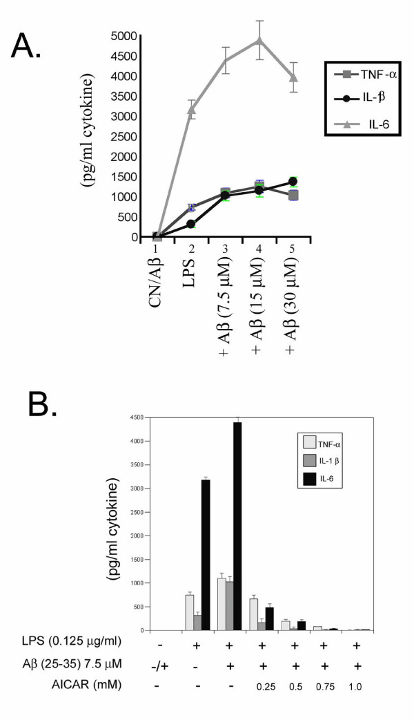
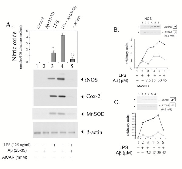
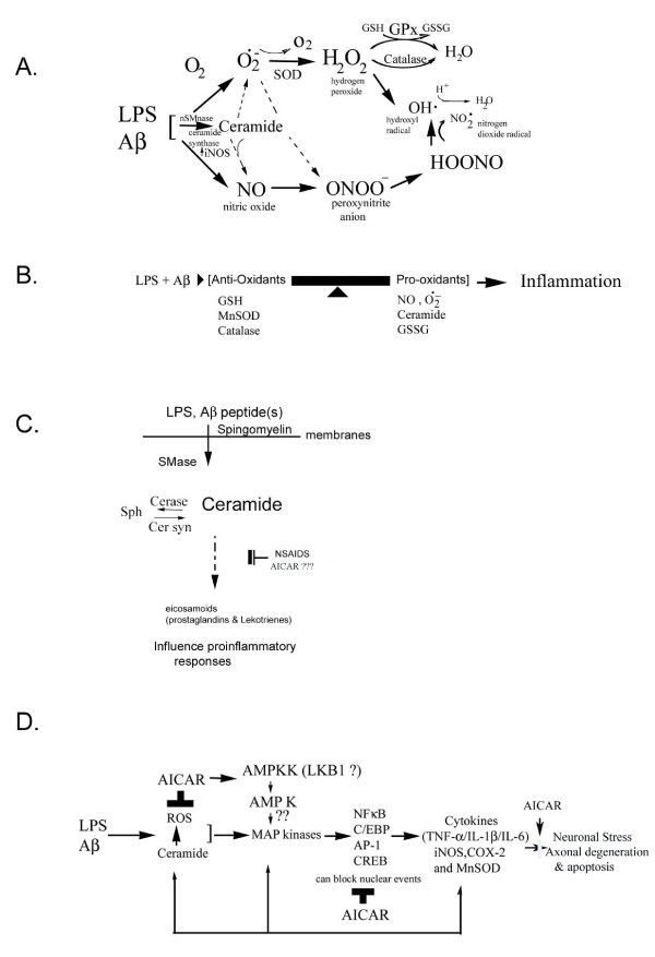
Background: Alzheimer's disease (AD) pathology shows characteristic 'plaques' rich in amyloid beta (Abeta) peptide deposits. Inflammatory process-related proteins such as pro-inflammatory cytokines have been detected in AD brain suggesting that an inflammatory immune reaction also plays a role in the pathogenesis of AD. Glial cells in culture respond to LPS and Abeta stimuli by upregulating the expression of cytokines TNF-alpha, IL-1beta, and IL-6, and also the expression of proinflammatory genes iNOS and COX-2. We have earlier reported that LPS/Abeta stimulation-induced ceramide and ROS generation leads to iNOS expression and nitric oxide production in glial cells. The present study was undertaken to investigate the neuroprotective function of AICAR (a potent activator of AMP-activated protein kinase) in blocking the pro-oxidant/proinflammatory responses induced in primary glial cultures treated with LPS and Abeta peptide.

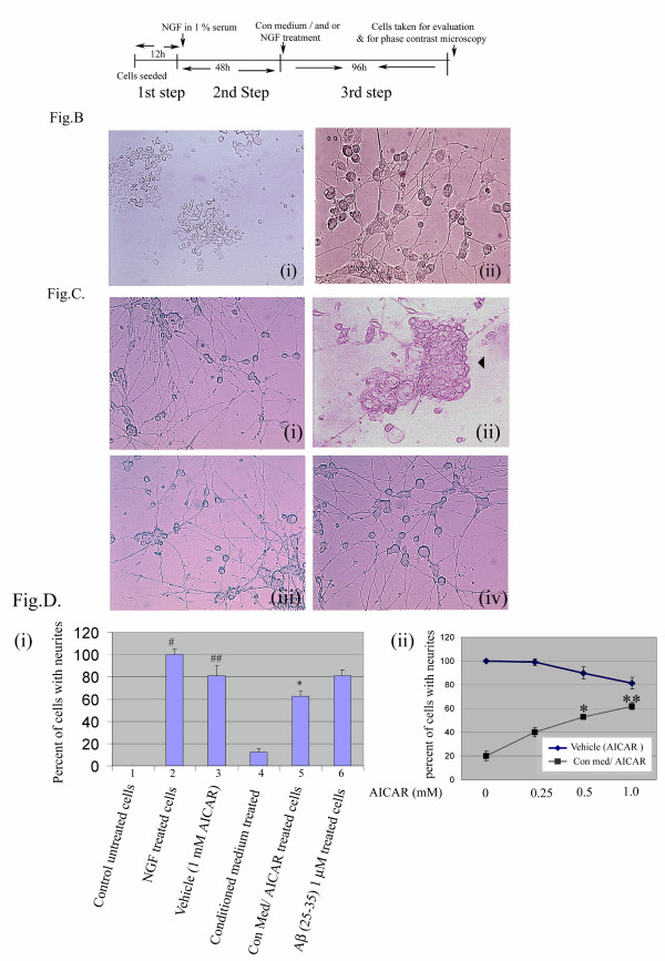
Methods: To test the anti-inflammatory/anti-oxidant functions of AICAR, we tested its inhibitory potential in blocking the expression of pro-inflammatory cytokines and iNOS, expression of COX-2, generation of ROS, and associated signaling following treatment of glial cells with LPS and Abeta peptide. We also investigated the neuroprotective effects of AICAR against the effects of cytokines and inflammatory mediators (released by the glia), in blocking neurite outgrowth inhibition, and in nerve growth factor-(NGF) induced neurite extension by PC-12 cells.

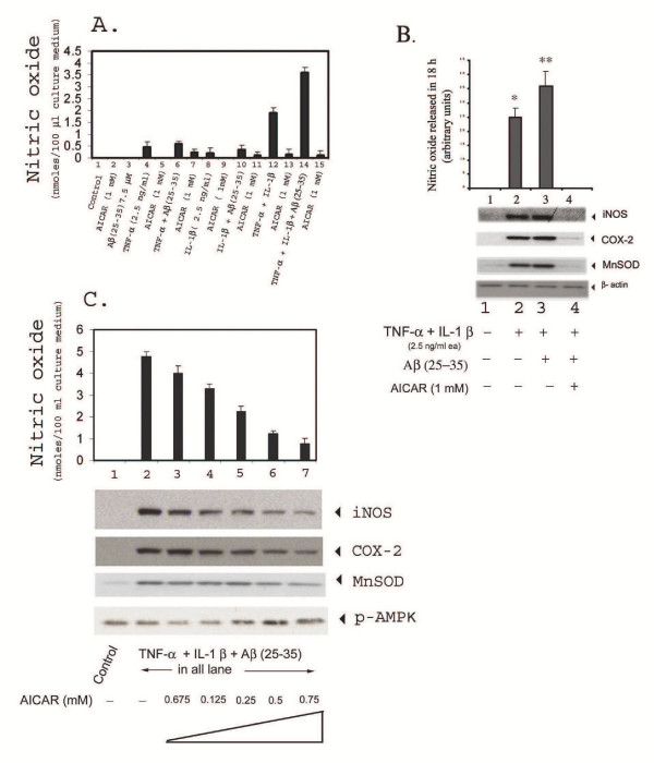
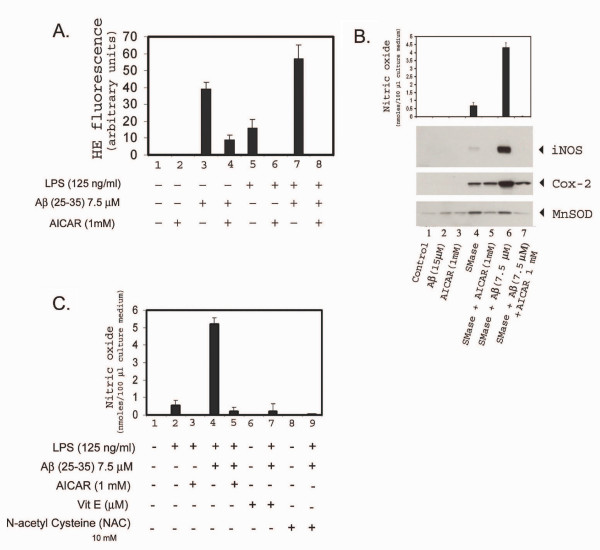
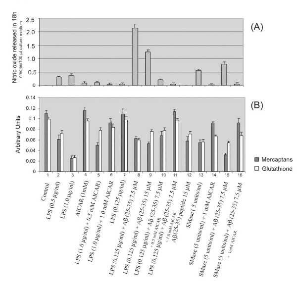
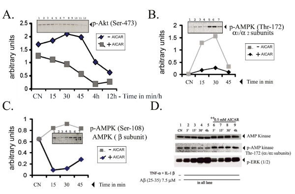
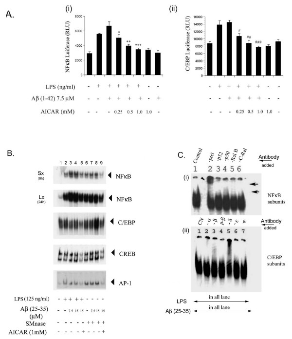
Results: AICAR blocked LPS/Abeta-induced inflammatory processes by blocking the expression of proinflammatory cytokine, iNOS, COX-2 and MnSOD genes, and by inhibition of ROS generation and depletion of glutathione in astroglial cells. AICAR also inhibited down-stream signaling leading to the regulation of transcriptional factors such as NFkappaB and C/EBP which are critical for the expression of iNOS, COX-2, MnSOD and cytokines (TNF-alpha/IL-1beta and IL-6). AICAR promoted NGF-induced neurite growth and reduced neurite outgrowth inhibition in PC-12 cells treated with astroglial conditioned medium.

Conclusion: The observed anti-inflammatory/anti-oxidant and neuroprotective functions of AICAR suggest it as a viable candidate for use in treatment of Alzheimer's disease.


Figures

Figure 1
Figure 1
AICAR inhibits LPS- and Aβ peptide-induced cytokine production. Astrocyte-enriched glial cells (mixed glial cells) were pre-incubated with different concentrations of AICAR (as indicated) for 4 h and were stimulated with 1 ng/ml LPS ± Aβ peptide (1–42) (15 μM) as shown. After 18 h of incubation, concentrations of NO, TNF-α, IL-1β, and IL-6 released into the culture medium were measured using ELISA (left panel figs. a, c, e and g). Alternatively the cells were harvested for RNA by extraction with Trizol (see methods) and the levels of mRNA for cytokines were measured (See right panel figs. b, d, f and h) by real time-PCR (RT-PCR). Data are expressed as the mean ± SD of three different experiments. *P < 0.001 was considered significant.
Figure 2
Figure 2
AICAR treatment inhibits LPS + Aβ-stimulated iNOS gene expression and nitric oxide release in glial cells. Cell cultures were pre-incubated with 1 mM AICAR following stimulation by LPS ± Aβ peptides (1–40) or (1–42) in concentrations as indicated. The corresponding reverse peptide (40–1) in lane 3 and 9 served as a positive control in this assay. The production of NO (top) and expression of iNOS, COX-2, and MnSOD was determined in cell lysates, 18 h following treatment, by immunoblot analysis (bottom). Experiments were performed in triplicate and data are means (±SEM). P < 0.05 compared to relative control value was considered significant. However, P value for histograms in lane 8 and 9 (* and **) not significant.
Figure 3
Figure 3
AICAR inhibits LPS- and Aβ (25–35) peptide-induced cytokine production in glial cells stimulated with 125 ng/ml LPS ± 7.5 μM Aβ peptide (25–35). After 18 h of incubation, concentrations of TNF-α, IL-1β, and IL-6 released into the culture medium were measured using ELISA. Fig. 3A. Shows dose-response curves, using LPS and various concentrations of Aβ (25–35) peptide in stimulating cytokine release in glial cells. Fig. 3B shows a dose-response inhibition of cytokine release with various concentrations of AICAR (0.25 to 1 mM) following stimulation with LPS + Aβ (25–35) peptide.
Figure 4
Figure 4
AICAR inhibits LPS- and Aβ- (25–35) induced expression of iNOS, COX-2, and MnSOD in astrocyte-enriched glial cells. Cells were preincubated with 1 mM AICAR for 4 h prior to treatment with either LPS or Aβ (25–35) in concentrations indicated earlier. After 18 h incubation, an aliquot of the medium was used for nitrite measurement as described under Methods. Data are mean ± SD of three different experiments. (A) Cell homogenates were used for western-immunoblot analysis of iNOS, COX-2 and MnSOD. Western immunoblots for iNOS (B) and MnSOD (C) upon treatment of glial cell cultures to LPS and to various concentrations of Aβ (25–35) either in the presence or absence of 0.5 mM AICAR (lane 1 is control, lane 2 LPS alone, and in lanes 3 to 6, Aβ was added to final concentrations of 7.5, 15, 30 or 45 μM, respectively). The protein bands were scanned on a densitometric scanner and represented as a graph (bottom). Experiments were performed in triplicate and data are means (±SEM). *P < 0.05 compared to relative control value was considered significant.
Figure 5
Figure 5
AICAR inhibits TNF-α-, and/or IL-1β- and/or Aβ- (25–35) stimulated iNOS expression and nitric oxide release in astrocytic cellcultures. Cells were pre-incubated with 1 mM AICAR prior to stimulation. Fig. A shows nitric oxide released into the medium upon treatment with cytokines +/- Aβ (25–35) with relevant controls. Figure B shows nitric oxide released into the medium and corresponding western-immunoblot for iNOS, COX-2, and MnSOD, after stimulation with cytokines (TNF-α + IL-1β ± Aβ peptide), either in the presence or absence of AICAR in concentrations used in figure A. Figure C shows a dose-dependent inhibition of nitric oxide production and expression of iNOS, COX-2 and MnSOD proteins, on stimulation with cytokines and Aβ and after pre-incubation with increasing amounts of AICAR, as shown. The increase in p-AMPK protein band (Thr-172) indicates activation of AMPK with increasing concentrations of AICAR.
Figure 6
Figure 6
AICAR down-regulates Aβ ± LPS- or sphingomyelinase-induced generation of reactive oxygen species (ROS) and expression of iNOS, COX-2 and MnSOD. Figure A shows LPS ± Aβ- (25–35) induced ROS generation in glial cell cultures. Pre-treatment of glial cells with AICAR inhibits LPS- and Aβ-mediated ROS generation. Cells were pre-incubated in the culture medium with 1 mM AICAR for 4 h prior to treatment with LPS (0.125 μg/ml) and/or 7.5 μM Aβ (25–35). ROS generation was measured by incubating the cells with the fluorescent dye Hydro Ethidene (HE) as described under Methods. Columns where AICAR was added have been shown diagramatically for brevity. Figure B: anti-oxidants (NAC and vitamin E) mediated inhibition of LPS, Aβ-stimulated nitric oxide release in glial cultures. Cells were pretreated with either AICAR (1 mM), vitamin E (20 μM) or N-acetyl cysteine (NAC) (10 mM) 4 h prior to stimulation with 125 ng/ml LPS and Aβ 7.5 μM (25–35). Nitric oxide released into the medium was measured as described under methods. Figure C shows AICAR mediated inhibition of both SMase- and Aβ-stimulated nitric oxide release (top) as well as the expression of iNOS, COX-2 and MnSOD in astrocyte enriched glial cultures (bottom). Cultures were pre-incubated with AICAR 4 h prior to stimulation with SMase (5 units/ml) ± 7.5 μM Aβ (25–35). Nitric oxide produced in the culture medium was measured using 'Greis reagent'. The cell lysates were western-immunoblotted for iNOS, COX-2 and MnSOD expression.
Figure 7
Figure 7
Effect of AICAR in normalization of LPS-, or SMase- and/or Aβ peptide- (25–35) induced decreases in cellular glutathione and total mercaptans (thiol group containing compounds). Figure A shows a histogram plot of NO released after treatment with LPS or SMase and/or Aβ peptide, either in the presence or absence of AICAR (1 mM). Figure B shows corresponding levels of glutathione (in light colored bars) and total mercaptans (dark colored bars).
Figure 8
Figure 8
AICAR inhibits LPS-and Aβ-induced activation of Pkb/Akt kinase activity, but activates AMP kinase activity, in astrocyte-enriched glial cell cultures. Cultures pretreated with AICAR or untreated cells were stimulated with LPS (0.125 μg/ml) and 7.5 μM Aβ for the indicated time periods following which cell homogenates were western-immunoblotted for phosphorylated forms of AMPK and Pkb/Akt. Figure 8A shows immunoblots for p-Pkb/Akt proteins. Samples (1 and 7) correspond to control, (2 and 8), (3 and 9), (4 and 10), (5 and 11), (6 and 12) correspond to cells treated with LPS + Aβ +/- 1 mM AICAR for 15 min, 30 min, 45 min, 4 h or 12 h respectively (as shown). Figure B shows phosphor-AMP K (Thr-172 of the α12 subunits) and Figure C to phosphorylated AMPK (Ser 108) of AMPK β subunit. In Figures B and C samples (1) control (2 and 5), (3 and 6) and (4 and 7) correspond to cells treated with LPS + Aβ peptide (for 15 min, 30 min or 60 min respectively) with or without AICAR pre-treatment. Fig D, shows AICAR-mediated inhibition of TNF-α/IL-1β- and Aβ-induced activation of ERK and activation of AMPK. Cells were pre-incubated with AICAR (1 mM) for 4 h prior to treatment with cytokine and Aβ (25–35). Cell homogenates were prepared at indicated time points and western immunoblotted for either phosphorylated or nonphosphorylated iso forms as shown.
Figure 9
Figure 9
(A) AICAR inhibits LPS- and Aβ-induced activation of NFκB, AP-1 CREB and C/EBP transcription factors. NFkB luciferase and C/EBP luciferase activities in glial cells transfected for NFkB luciferase (i) or C/EBP luciferase following stimulation with LPS and Aβ (1–42) peptide (figure A) were measured as described under legends to figure 1. Experiments were performed in triplicate and data are expressed as mean ± SEM. *P < 0.05 compared to relative control value was considered significant. AICAR inhibited the LPS- or SMase- and Aβ-induced NFκB, C/EBP, CREB and AP-1 binding activity as seen by EMSA (figure B). EMSA was carried out using the nuclear extracts prepared from astrocyte-enriched glial cells after treatment with LPS or SMase and Aβ for 1 h either in the presence or absence of AICAR. In case of EMSA for NFκB, for clarity, the dried gel was exposed for autoradiography either for longer (24 h) or for shorter (6 h) periods. The top shows the picture of the autoradiogram of the shorter exposure time (Sx) and the bottom shows the longer exposure time (Lx). In figure B(i) polyclonal IgGs specific for NFκB subunits -p65, p52, p50, RelB or cRel were used for supershift experiments with nuclear extracts from LPS, Aβ-treated (1 h) glial cells for binding to γ-32P-labeled NFκB oligomer. Note the supershifted complexes in lanes 2, 4 and 5 (correspond to -p65, p50 and -RelB proteins). In figure (ii) lanes 2–7, polyclonal IgGs specific for C/EBP α, β, p-β, δ and ε were used in supershift experiments with nuclear extracts from LPS, Aβ-treated (1 h) glial cells for binding to γ-32P-labeled C/EBP oligomer. ε antibodies from two different stocks were tested (in lanes 6 and 7). Note the supershifted complexes in lanes 2, 3 and 5 corresponding to -α, β and -δ subunits of C/EBP.
Figure 10
Figure 10
AICAR promotes PC-12 cell neurite extension following stimulation with NGF in the presence of glial conditioned medium. Figure A shows the scheme of treatment using glial cell-conditioned medium (see Methods) either in the presence or absence of AICAR (1 mM). Figures B shows phase contrast micrographs of PC-12 cells of control untreated cells and NGF differentiated PC-12 cells. Where figure B(i) shows control untreated cells (40× magnification) and treated with NGF (50 ng/ml) form neurites reminiscent of axonal network in neurons as shown in figure B (ii) at 40× magnification. AICAR protection against glial conditioned medium is demonstrated in figure 10C where figure (i) is vehicle (control) NGF + 1 mM AICAR treatment. Inhibition of NGF-induced neurite outgrowth observed on treatment with glia-conditioned medium (from LPS, Aβ treatment) showing aggregation of cells (ii) and its reversal in AICAR pre-treated and NGF- and conditioned medium-challenged cells (iii), (all at 20× magnification). Aβ (25–35) (1 μM) treatment was performed as one of the controls (iv) 20× magnification. Experiments were carried out in triplicate. Figure D shows a histogram plot of neurite lengths greater than or equal to 100 μm in the above experiment in vehicle-treated, condition medium-treated, AICAR-treated or Aβ- (25–35) treated PC-12 cells. Data shown are mean ± SEM. # p < 0.5 was considered significant. Figure E shows the effects of different concentrations of AICAR in the presence of fixed amount of conditioned medium on neurite outgrowth. AICAR was tried at three different concentrations against condition medium challenged cells. Values shown are relative percentage values. The neurite outgrowth was recorded after 96 h of NGF stimulation. Quantitative analysis of neurite outgrowth is from ~300 cells. The data was plotted as mean ± SD from three experiments. *p < 0.5 was considered significant. P values for Conditioned medium/AICAR treated samples * and ** significant.
Figure 11
Figure 11
Figure A: Possible anti-oxidant mechanisms involved in the LPS, Aβ-induced ceramide generation leading to superoxide anion formation, nitric oxide release and peroxynitrite generation. Figure B: inflammation is possibly triggered as a result of imbalance in the radical generating systems and radical scavenger systems creating an oxidative stress, thus leading to the formation of nitric oxide and superoxide anion generation, and thereby depleting cellular anti-oxidants. Figure C. Putative role of ceramide in eicosanoid synthesis. Ceramide generated as a result of LPS- and Aβ-peptide induced SMase activation, leads to the release of eicosanoids. Eicosanoids (leukotrienes and prostaglandins) thus generated may perhaps potentially enhance or ameliorate the cytokine-induced pro-inflammatory responses or vice versa. Non steroidal anti-inflammatory drugs (NSAIDs) as well as COX inhibitors block these responses. Figure D. Overall scheme in the LPS, Aβ-induced pro-inflammatory signaling cascade involving cytokine release thereby leading to the expression of iNOS, COX-2 and MnSOD. The anti-inflammatory effect of AICAR is perhaps a result of its multiple regulatory roles. However, AICAR blockade of ROS generation keeps the redox balance in check, thereby inhibiting the inflammatory signaling cascade.

Similar articles

Cited by

References

    1. Selkoe DJ. Alzheimer's disease results from the cerebral accumulation and cytotoxicity of amyloid beta-protein. J Alzheimers Dis. 2001;3:75–80. - PubMed
    1. Abramov AY, Canevari L, Duchen MR. Beta-amyloid peptides induce mitochondrial dysfunction and oxidative stress in astrocytes and death of neurons through activation of NADPH oxidase. J Neurosci. 2004;24:565–575. doi: 10.1523/JNEUROSCI.4042-03.2004. - DOI - PMC - PubMed
    1. McGeer EG, McGeer PL. Inflammatory processes in Alzheimer's disease. Prog Neuropsychopharmacol Biol Psychiatry. 2003;27:741–749. doi: 10.1016/S0278-5846(03)00124-6. - DOI - PubMed
    1. Akiyama H, Barger S, Barnum S, Bradt B, Bauer J, Cole GM, Cooper NR, Eikelenboom P, Emmerling M, Fiebich BL, Finch CE, Frautschy S, Griffin WS, Hampel H, Hull M, Landreth G, Lue L, Mrak R, Mackenzie IR, McGeer PL, O'Banion MK, Pachter J, Pasinetti G, Plata-Salaman C, Rogers J, Rydel R, Shen Y, Streit W, Strohmeyer R, Tooyoma I, Van Muiswinkel FL, Veerhuis R, Walker D, Webster S, Wegrzyniak B, Wenk G, Wyss-Coray T. Inflammation and Alzheimer's disease. Neurobiol Aging. 2000;21:383–421. doi: 10.1016/S0197-4580(00)00124-X. - DOI - PMC - PubMed
    1. Tsukamoto E, Hashimoto Y, Kanekura K, Niikura T, Aiso S, Nishimoto I. Characterization of the toxic mechanism triggered by Alzheimer's amyloid-beta peptides via p75 neurotrophin receptor in neuronal hybrid cells. J Neurosci Res. 2003;73:627–636. doi: 10.1002/jnr.10703. - DOI - PubMed