

Autophagy in chronically ischemic myocardium
- PMID: 16174725
- PMCID: PMC1224362
- DOI: 10.1073/pnas.0506843102
Autophagy in chronically ischemic myocardium
Abstract
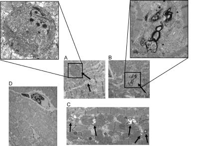
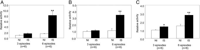
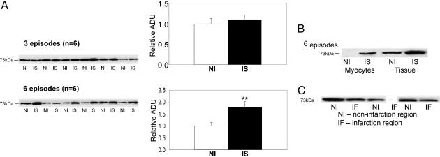
We tested the hypothesis that chronically ischemic (IS) myocardium induces autophagy, a cellular degradation process responsible for the turnover of unnecessary or dysfunctional organelles and cytoplasmic proteins, which could protect against the consequences of further ischemia. Chronically instrumented pigs were studied with repetitive myocardial ischemia produced by one, three, or six episodes of 90 min of coronary stenosis (30% reduction in baseline coronary flow followed by reperfusion every 12 h) with the non-IS region as control. In this model, wall thickening in the IS region was chronically depressed by approximately 37%. Using a nonbiased proteomic approach combining 2D gel electrophoresis with in-gel proteolysis, peptide mapping by MS, and sequence database searches for protein identification, we demonstrated increased expression of cathepsin D, a protein known to mediate autophagy. Additional autophagic proteins, cathepsin B, heat shock cognate protein Hsc73 (a key protein marker for chaperone-mediated autophagy), beclin 1 (a mammalian autophagy gene), and the processed form of microtubule-associated protein 1 light chain 3 (a marker for autophagosomes), were also increased. These changes, not evident after one episode, began to appear after two or three episodes and were most marked after six episodes of ischemia, when EM demonstrated autophagic vacuoles in chronically IS myocytes. Conversely, apoptosis, which was most marked after three episodes, decreased strikingly after six episodes, when autophagy had increased. Immunohistochemistry staining for cathepsin B was more intense in areas where apoptosis was absent. Thus, autophagy, triggered by ischemia, could be a homeostatic mechanism, by which apoptosis is inhibited and the deleterious effects of chronic ischemia are limited.
Figures


References
-
- Melendez, A., Talloczy, Z., Seaman, M., Eskelinen, E. L., Hall, D. H. & Levine, B. (2003) Science 301, 1387–1391. - PubMed
-
- Stromhaug, P. E. & Klionsky, D. J. (2001) Traffic 2, 524–531. - PubMed
-
- Huang, W. P. & Klionsky, D. J. (2002) Cell Struct. Funct. 27, 409–420. - PubMed
-
- Ohsumi, Y. (2001) Nat. Rev. Mol. Cell. Biol. 2, 211–216. - PubMed
-
- Otto, G. P., Wu, M. Y., Kazgan, N., Anderson, O. R. & Kessin, R. H. (2003) J. Biol. Chem. 278, 17636–17645. - PubMed
Publication types
MeSH terms
Substances
Grants and funding
- HL65183/HL/NHLBI NIH HHS/United States
- 2R01HL33107/HL/NHLBI NIH HHS/United States
- HL65182/HL/NHLBI NIH HHS/United States
- K12 HD001457/HD/NICHD NIH HHS/United States
- HD01457/HD/NICHD NIH HHS/United States
- 2R01AG14121/AG/NIA NIH HHS/United States
- 2P01HL59139/HL/NHLBI NIH HHS/United States
- R01 HL033107/HL/NHLBI NIH HHS/United States
- 1P01HL69020/HL/NHLBI NIH HHS/United States
- P01 HL069020/HL/NHLBI NIH HHS/United States
- R01 HL065182/HL/NHLBI NIH HHS/United States
- P01 HL059139/HL/NHLBI NIH HHS/United States
- R01 AG023137/AG/NIA NIH HHS/United States
- R01 AG014121/AG/NIA NIH HHS/United States
- AG023137/AG/NIA NIH HHS/United States
LinkOut - more resources
Full Text Sources
Research Materials
Miscellaneous
