Dissection of the carbohydrate specificity of the broadly neutralizing anti-HIV-1 antibody 2G12
- PMID: 16174734
- PMCID: PMC1224641
- DOI: 10.1073/pnas.0505763102
Dissection of the carbohydrate specificity of the broadly neutralizing anti-HIV-1 antibody 2G12
Abstract
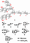
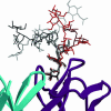
Human antibody 2G12 neutralizes a broad range of HIV-1 isolates. Hence, molecular characterization of its epitope, which corresponds to a conserved cluster of oligomannoses on the viral envelope glycoprotein gp120, is a high priority in HIV vaccine design. A prior crystal structure of 2G12 in complex with Man(9)GlcNAc(2) highlighted the central importance of the D1 arm in antibody binding. To characterize the specificity of 2G12 more precisely, we performed solution-phase ELISA, carbohydrate microarray analysis, and cocrystallized Fab 2G12 with four different oligomannose derivatives (Man(4), Man(5), Man(7), and Man(8)) that compete with gp120 for binding to 2G12. Our combined studies reveal that 2G12 is capable of binding both the D1 and D3 arms of the Man(9)GlcNAc(2) moiety, which would provide more flexibility to make the required multivalent interactions between the antibody and the gp120 oligomannose cluster than thought previously. These results have important consequences for the design of immunogens to elicit 2G12-like neutralizing antibodies as a component of an HIV vaccine.
Figures


Similar articles
-
A solution NMR study of the interactions of oligomannosides and the anti-HIV-1 2G12 antibody reveals distinct binding modes for branched ligands.Chemistry. 2011 Feb 1;17(5):1547-60. doi: 10.1002/chem.201002519. Epub 2011 Jan 5. Chemistry. 2011. PMID: 21268157
-
Binding of high-mannose-type oligosaccharides and synthetic oligomannose clusters to human antibody 2G12: implications for HIV-1 vaccine design.Chem Biol. 2004 Jan;11(1):127-34. doi: 10.1016/j.chembiol.2003.12.020. Chem Biol. 2004. PMID: 15113002
-
A glycoconjugate antigen based on the recognition motif of a broadly neutralizing human immunodeficiency virus antibody, 2G12, is immunogenic but elicits antibodies unable to bind to the self glycans of gp120.J Virol. 2008 Jul;82(13):6359-68. doi: 10.1128/JVI.00293-08. Epub 2008 Apr 23. J Virol. 2008. PMID: 18434393 Free PMC article.
-
Antibody responses to the HIV-1 envelope high mannose patch.Adv Immunol. 2019;143:11-73. doi: 10.1016/bs.ai.2019.08.002. Epub 2019 Sep 11. Adv Immunol. 2019. PMID: 31607367 Free PMC article. Review.
-
GP120: target for neutralizing HIV-1 antibodies.Annu Rev Immunol. 2006;24:739-69. doi: 10.1146/annurev.immunol.24.021605.090557. Annu Rev Immunol. 2006. PMID: 16551265 Review.
Cited by
-
Antibody WN1 222-5 mimics Toll-like receptor 4 binding in the recognition of LPS.Proc Natl Acad Sci U S A. 2012 Dec 18;109(51):20877-82. doi: 10.1073/pnas.1209253109. Epub 2012 Nov 26. Proc Natl Acad Sci U S A. 2012. PMID: 23184990 Free PMC article.
-
Conference report: "Functional Glycomics in HIV Type 1 Vaccine Design" workshop report, Bethesda, Maryland, April 30-May 1, 2012.AIDS Res Hum Retroviruses. 2013 Nov;29(11):1407-17. doi: 10.1089/AID.2013.0102. Epub 2013 Jul 19. AIDS Res Hum Retroviruses. 2013. PMID: 23767872 Free PMC article.
-
De novo asymmetric synthesis of all-D-, all-L-, and D-/L-oligosaccharides using atom-less protecting groups.J Am Chem Soc. 2012 Jul 25;134(29):11952-5. doi: 10.1021/ja305321e. Epub 2012 Jul 16. J Am Chem Soc. 2012. PMID: 22780712 Free PMC article.
-
Targeting the glycans of glycoproteins: a novel paradigm for antiviral therapy.Nat Rev Microbiol. 2007 Aug;5(8):583-97. doi: 10.1038/nrmicro1707. Nat Rev Microbiol. 2007. PMID: 17632570 Free PMC article. Review.
-
Yeast-elicited cross-reactive antibodies to HIV Env glycans efficiently neutralize virions expressing exclusively high-mannose N-linked glycans.J Virol. 2011 Jan;85(1):470-80. doi: 10.1128/JVI.01349-10. Epub 2010 Oct 20. J Virol. 2011. PMID: 20962094 Free PMC article.
References
-
- Letvin, N. L. & Walker, B. D. (2003) Nat. Med. 9, 861-866. - PubMed
-
- McMichael, A. J. & Hanke, T. (2003) Nat. Med. 9, 874-880. - PubMed
-
- Burton, D. R., Desrosiers, R. C., Doms, R. W., Koff, W. C., Kwong, P. D., Moore, J. P., Nabel, G. J., Sodroski, J., Wilson, I. A. & Wyatt, R. T. (2004) Nat. Immunol. 5, 233-236. - PubMed
Publication types
MeSH terms
Substances
Associated data
- Actions
- Actions
- Actions
- Actions
Grants and funding
LinkOut - more resources
Full Text Sources
Other Literature Sources

