. 2005 Sep 20;102(38):13461-6.
doi: 10.1073/pnas.0503689102. Epub 2005 Sep 7.

Neuronal sorting protein-related receptor sorLA/LR11 regulates processing of the amyloid precursor protein

Affiliations

Neuronal sorting protein-related receptor sorLA/LR11 regulates processing of the amyloid precursor protein

Olav M Andersen et al. Proc Natl Acad Sci U S A. .

Abstract

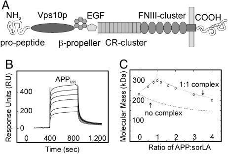
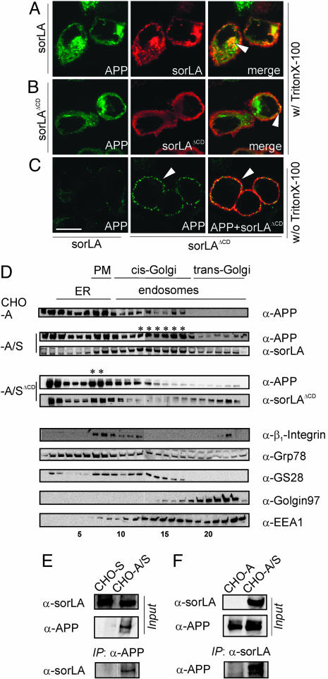
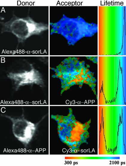
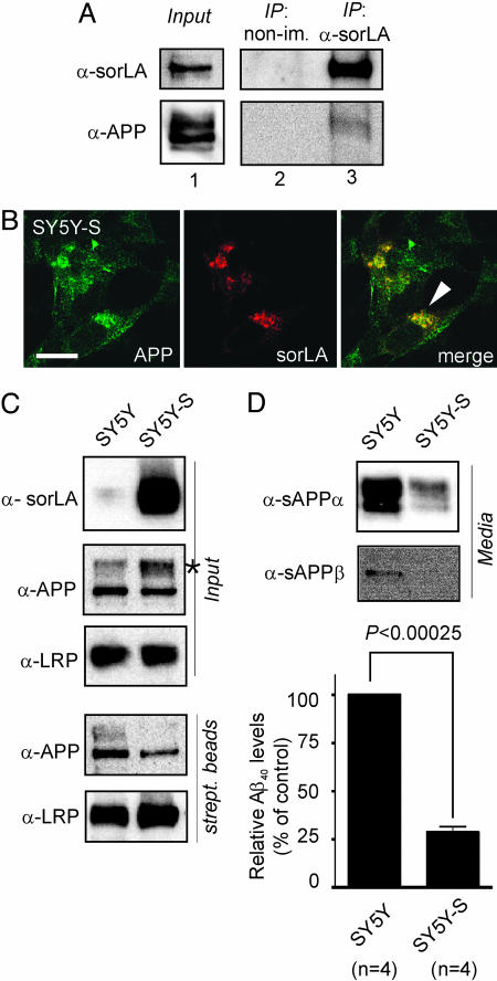
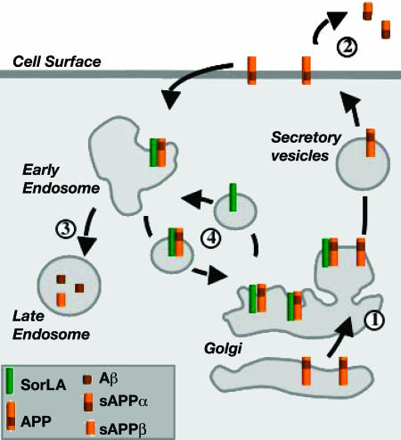
sorLA (Sorting protein-related receptor) is a type-1 membrane protein of unknown function that is expressed in neurons. Its homology to sorting receptors that shuttle between the plasma membrane, endosomes, and the Golgi suggests a related function in neuronal trafficking processes. Because expression of sorLA is reduced in the brain of patients with Alzheimer's disease (AD), we tested involvement of this receptor in intracellular transport and processing of the amyloid precursor protein (APP) to the amyloid beta-peptide (Abeta), the principal component of senile plaques. We demonstrate that sorLA interacts with APP in vitro and in living cells and that both proteins colocalize in endosomal and Golgi compartments. Overexpression of sorLA in neurons causes redistribution of APP to the Golgi and decreased processing to Abeta, whereas ablation of sorLA expression in knockout mice results in increased levels of Abeta in the brain similar to the situation in AD patients. Thus, sorLA acts as a sorting receptor that protects APP from processing into Abeta and thereby reduces the burden of amyloidogenic peptide formation. Consequently, reduced receptor expression in the human brain may increase Abeta production and plaque formation and promote spontaneous AD.


Figures

Fig. 1.
Fig. 1.
APP and sorLA interact in vitro.(A) The structural elements of sorLA are depicted, including (from amino to carboxyl terminus) the propeptide, vacuolar protein sorting 10 protein (Vps10p) domain, β-propeller, epidermal growth factor repeat (EGF), and clusters of complement-type repeats (CR) and fibronectin type III domains (FNIII). (B and C) The sorLA ectodomain used for surface plasmon resonance (SPR) (B) or molecular mass analysis (C) encompassed all elements amino-terminal to the membrane anchor. (B) SPR analysis of binding of a concentration series (0.1, 0.2, 0.5, 1, 2, and 5 μM) of APP695 to sorLA immobilized on the sensor chip (KD = 200 nM). (C) Molecular-weight plot of APP-sorLA complex formation by analytic ultracentrifugation. The sorLA concentration was kept at 0.1 μM, whereas the APP695 concentration varied to give the indicated APP:sorLA input ratios. Circles indicate the actual data points of the observed average molecular masses in the protein mixture at a given APP:sorLA ratio. Lines represent theoretical graphs to be expected for a perfect 1:1 complex (dotted) or no complex formation (dashed).
Fig. 2.
Fig. 2.
SorLA affects trafficking of APP in CHO cells. (A-C) Detection of APP695 and sorLA in CHO cells by using confocal immunofluorescence microscopy. (A) In permeabilized cells (with Triton X-100), APP695 and wild-type sorLA signals colocalize to intracellular vesicular structures and to the perinuclear region (arrowhead). (B) In stable transfectants coexpressing APP695 and sorLAΔCD, the sorLAΔCD signal is confined to the cell surface, where it colocalizes with APP (arrowhead). (C) In nonpermeabilized cells (without Triton X-100), more APP695 is detected on the surface in cells that express sorLAΔCD (Center, arrowhead), where it colocalizes with the mutant receptor (Right, arrowhead) compared with cells expressing wild-type sorLA (Left). (Scale bar: 10 μm.) (D) Subcellular fractionation of cells expressing APP695 (CHO-A) or APP695 together with wild-type sorLA (CHO-A/S) or sorLAΔCD (CHO-A/SΔCD). Immunodetection of APP and sorLA, as well as marker proteins β1-integrin (plasma membrane, PM), Grp78 (endoplasmic reticulum, ER), GS28 (cis-Golgi), Golgin97 (trans-Golgi network), and EEA1 (early endosomes) in the fractions is depicted. Asterisks mark coaccumulation of APP and sorLA in Golgi or in plasma membrane compartments of CHO-A/S and CHO-A/SΔCD cells, respectively. (E and F) Cells expressing sorLA (CHO-S), APP695 (CHO-A), or both (CHO-A/S) were treated with membrane-permeable cross-linker and proteins immunoprecipitated by using anti-APP (E) or anti-sorLA (F) antibodies. Western blots labeled “Input” show sorLA and APP in total cell extracts before immunoprecipitation. Western blots labeled “IP” demonstrate coimmunoprecipitation of sorLA in anti-APP (E) and APP in anti-sorLA (F) precipitates.
Fig. 3.
Fig. 3.
FLIM of APP695 and sorLA interaction in N2A cells. Shown are intensity images of sorLA (A and B) and APP695 (C) immunocytochemistry using donor fluorophore Alexa Fluor 488-conjugated antibodies (Left), pseudocolored FLIM images (Center), and lifetime histograms (Right), indicating shortening of lifetime from blue to orange/red in the absence (A) or presence (B and C) of acceptor fluorophore Cy3. (A) Cells stained for the amino-terminal domain of sorLA with donor fluorophore Alexa Fluor 488 in the absence of acceptor (lifetime of 2,007 ± 12 psec). (B) Cells stained for sorLA with donor fluorophore Alexa Fluor 488 in the presence of acceptor Cy3 on the ectodomain of APP695 (lifetime reduced to 1,365 ± 139 psec). (C) Cells stained for APP with donor Alexa Fluor 488 in the presence of acceptor Cy3 on sorLA (lifetime reduced to 1,481 ± 228 psec).
Fig. 4.
Fig. 4.
SorLA alters processing of APP in SH-SY5Y cells. (A) Nontransfected SH-SY5Y cells were treated with membrane-permeable cross-linker, and proteins were immunoprecipitated by using anti-sorLA antibodies (IP: α-sorLA) or nonimmune serum (IP: non-im.). Lane 1 shows Western blots of endogenous sorLA and APP expression in cell extracts before immunoprecipitation. Also shown are Western blots for sorLA and APP in nonimmune (lane 2) or anti-sorLA (lane 3) immunoprecipitates. (B) Immunodetection of endogenous APP and sorLA in SH-SY5Y cells transfected with a sorLA expression construct (SY5Y-S) indicating colocalization in the perinuclear region (arrowhead). (Scale bar: 10 μm.) (C) Surface biotinylation demonstrating decreased surface localization of APP in cells overexpressing sorLA. Blots labeled “Input” depict sorLA, APP, and LRP expression in cell extracts from parental (SY5Y) and sorLA-transfected (SY5Y-S) neurons. The asterisk indicates accumulation of mature APP in the presence of sorLA. Western blots labeled “strept. beads” show biotinylated APP and LRP in precipitates from streptavidin beads. Overexpression of sorLA reduces cell surface exposure of APP (35.8% of normal) but not of LRP in SY5Y-S compared with SY5Y cells. (D) Levels of sAPPα, sAPPβ, and Aβ40 in medium from SY5Y and SY5Y-S cells as determined by Western blot or ELISA. Aβ40 in SY5Y-S is reduced to 28.6 ± 3.0% (with nontransfected SY5Y levels set at 100%).
Fig. 5.
Fig. 5.
APP metabolism in AD patients and in sorLA-deficient mice. (A and B) SorLA levels are significantly reduced in the frontal cortex gray matter of 10 AD patients compared with 7 healthy subjects as shown by densitometric scanning (B) of Western blots, such as those shown in A. As a control, the levels of neuronal proteins LRP and synaptophysin were documented. n, number of patients. (C) Immunofluorescence detection of endogenous APP and sorLA in primary neurons from wild-type mice indicating partial colocalization in vesicular structures (arrowhead). (D-F) Immunodetection of sorLA (D), APP (E), and Aβ (F) in the frontal cortex of wild-type (+/+) and sorLA-deficient (-/-) mice. Representative data from experiments in three sorLA-/- mice and three control mice are shown. (Scale bar: 100 μm.) (G) Loss of sorLA expression in sorLA-/- mice (α-sorLA) did not affect the total levels of APP (α-APP) but increased processing into soluble APP (α-sAPP) as shown by Western blot analysis of brain homogenates. (H) Quantification of the increase in murine Aβ40 and Aβ42 in cortex extracts from 10-month-old sorLA-/- mice (138.6 ± 12.3% SEM and 135.6 ± 11.1% SEM, respectively) compared with wild-type controls (mean value set at 100%) using ELISA. P values were determined by equal-variance t test. n, number of mice.
Fig. 6.
Fig. 6.
Proposed role for sorLA in APP processing. Newly synthesized APP molecules traverse the Golgi (“1”) to the plasma membrane, where some are cleaved to sAPPα (“2”). Nonprocessed precursors endocytose from the cell surface into late endosomal compartments for processing into sAPPβ and Aβ (“3”). SorLA acts as a sorting receptor that traps APP in the Golgi (“1”), reducing the amount of precursors that reach the cell surface for processing. In addition, sorLA may also shuttle APP from early endosomes back to the Golgi, further reducing the extent of Aβ production in late endosomes (“4”). Consistent with this model, overexpression of sorLA in cultured cells further reduces transition of APP to the cell surface and suppresses processing into sAPPα and Aβ, whereas loss of sorLA expression in AD patients and in knockout mice results in accelerated trafficking of APP into processing pathways and in increased production of sAPPα and Aβ.

Similar articles

Cited by

References

    1. Hermans-Borgmeyer, I., Hampe, W., Schinke, B., Methner, A., Nykjaer, A., Susens, U., Fenger, U., Herbarth, B. & Schaller, H. C. (1998) Mech. Dev. 70, 65-76. - PubMed
    1. Motoi, Y., Aizawa, T., Haga, S., Nakamura, S., Namba, Y. & Ikeda, K. (1999) Brain Res. 833, 209-215. - PubMed
    1. Jacobsen, L., Madsen, P., Moestrup, S. K., Lund, A. H., Tommerup, N., Nykjaer, A., Sottrup-Jensen, L., Gliemann, J. & Petersen, C. M. (1996) J. Biol. Chem. 271, 31379-31383. - PubMed
    1. Yamazaki, H., Bujo, H., Kusunoki, J., Seimiya, K., Kanaki, T., Morisaki, N., Schneider, W. J. & Saito, Y. (1996) J. Biol. Chem. 271, 24761-24768. - PubMed
    1. Marcusson, E. G., Horazdovsky, B. F., Cereghino, J. L., Gharakhanian, E. & Emr, S. D. (1994) Cell 77, 579-586. - PubMed

Publication types

MeSH terms