

Cooperative interaction of Angiopoietin-like proteins 1 and 2 in zebrafish vascular development
- PMID: 16174743
- PMCID: PMC1224617
- DOI: 10.1073/pnas.0501902102
Cooperative interaction of Angiopoietin-like proteins 1 and 2 in zebrafish vascular development
Abstract
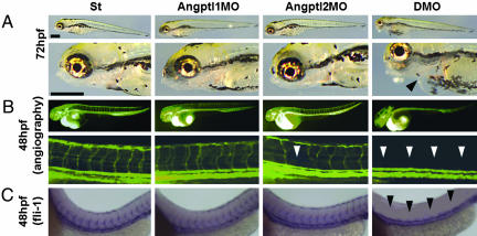
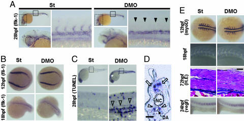
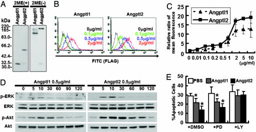
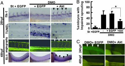
Angiopoietin-like protein (Angptl) 1 and Angptl2, which are considered orphan ligands, are highly homologous, particularly in the fibrinogen-like domain containing the putative receptor binding site. This similarity suggests potentially cooperative functions between the two proteins. In this report, the function of Angptl1 and Angptl2 is analyzed by using morpholino antisense technology in zebrafish. Knockdown of both Angptl1 and Angptl2 produced severe vascular defects due to increased apoptosis of endothelial cells at the sprouting stage. In vitro studies showed that Angptl1 and Angptl2 have antiapoptotic activities through the phosphatidylinositol 3-kinase/Akt pathway, and coinjection of constitutively active Akt/protein kinase B mRNA rescued impaired vascular development seen in double knockdown embryos. These results provide a physiological demonstration of the cooperative interaction of Angptl1 and Angptl2 in endothelial cells through phosphatidylinositol 3-kinase/Akt mediated antiapoptotic activities.
Figures
Similar articles
-
Angiopoietin-like proteins stimulate HSPC development through interaction with notch receptor signaling.Elife. 2015 Feb 25;4:e05544. doi: 10.7554/eLife.05544. Elife. 2015. PMID: 25714926 Free PMC article.
-
Angiopoietin-like protein 2 regulates endothelial colony forming cell vasculogenesis.Angiogenesis. 2014 Jul;17(3):675-83. doi: 10.1007/s10456-014-9423-8. Epub 2014 Feb 23. Angiogenesis. 2014. PMID: 24563071 Free PMC article.
-
Essential role of Apelin signaling during lymphatic development in zebrafish.Arterioscler Thromb Vasc Biol. 2014 Feb;34(2):338-45. doi: 10.1161/ATVBAHA.113.302785. Epub 2013 Dec 5. Arterioscler Thromb Vasc Biol. 2014. PMID: 24311379 Free PMC article.
-
The Two Faces of Angiopoietin-Like Protein 2 in Cancer.Cancer Sci. 2025 Mar;116(3):592-599. doi: 10.1111/cas.16434. Epub 2024 Dec 17. Cancer Sci. 2025. PMID: 39686837 Free PMC article. Review.
-
Angiopoietin-like proteins--potential therapeutic targets for metabolic syndrome and cardiovascular disease.Circ J. 2009 Dec;73(12):2192-7. doi: 10.1253/circj.cj-09-0710. Epub 2009 Oct 29. Circ J. 2009. PMID: 19875897 Review.
Cited by
-
Positional plasticity in regenerating Amybstoma mexicanum limbs is associated with cell proliferation and pathways of cellular differentiation.BMC Dev Biol. 2015 Nov 23;15:45. doi: 10.1186/s12861-015-0095-4. BMC Dev Biol. 2015. PMID: 26597593 Free PMC article.
-
Angiopoietin-like 2 is essential to aortic valve development in mice.Commun Biol. 2022 Nov 21;5(1):1277. doi: 10.1038/s42003-022-04243-6. Commun Biol. 2022. PMID: 36414704 Free PMC article.
-
Extracellular matrix-bound angiopoietin-like 4 inhibits endothelial cell adhesion, migration, and sprouting and alters actin cytoskeleton.Circ Res. 2006 Nov 24;99(11):1207-15. doi: 10.1161/01.RES.0000250758.63358.91. Epub 2006 Oct 26. Circ Res. 2006. PMID: 17068295 Free PMC article.
-
Synoviocyte-derived angiopoietin-like protein 2 contributes to synovial chronic inflammation in rheumatoid arthritis.Am J Pathol. 2010 May;176(5):2309-19. doi: 10.2353/ajpath.2010.090865. Epub 2010 Mar 19. Am J Pathol. 2010. PMID: 20304962 Free PMC article.
-
Upregulation of podocyte-secreted angiopoietin-like-4 in diabetic nephropathy.Endocrine. 2015 Jun;49(2):373-84. doi: 10.1007/s12020-014-0486-5. Epub 2014 Nov 26. Endocrine. 2015. PMID: 25424436
References
-
- Suri, C., Jones, P. F., Patan, S., Bartunkova, S., Maisonpierre, P., Davis, S., Sato, T. N. & Yancopoulos, G. D. (1996) Cell 87, 1171-1180. - PubMed
-
- Sato, T. N., Tozawa, Y., Deutsch, U., Wolburg-Buchholz, K., Fujiwara, Y., Gendron-Maguire, M., Grigley, T., Wolburg, H., Risau, W. & Qin, Y. (1995) Nature 376, 70-74. - PubMed
-
- Koblizek, T. I., Weiss, C., Yancopoulos, G. D., Deutsch, U. & Risau, W. (1998) Curr. Biol. 8, 529-532. - PubMed
-
- Witzenbichler, B., Maisonpierre, P. C., Jones, P., Yancopoulos, G. D. & Isner, J. M. (1998) J. Biol. Chem. 273, 18514-18521. - PubMed
Publication types
MeSH terms
Substances
LinkOut - more resources
Full Text Sources
Other Literature Sources
Molecular Biology Databases
