. 2005 Aug;15(3):207-13.
doi: 10.1055/s-2005-872596.

Extracranial-to-intracranial bypass using radial artery grafting for complex skull base tumors: technical note

Affiliations

Extracranial-to-intracranial bypass using radial artery grafting for complex skull base tumors: technical note

Saleem I Abdulrauf. Skull Base. 2005 Aug.

Abstract

The management of complex skull base tumors that incorporate large intracranial vessels poses challenging questions. Patients who fail initial surgical resection and adjunctive therapies (i.e., radiosurgery) who present with tumor regrowth may be candidates for parent vessel occlusion and total tumor resection in combination with extracranial-to-intracranial (EC-IC) bypass to augment the sacrificed vessel territory. In this technical report, we delineate the surgical technique of performing an EC-IC bypass using a radial artery graft. Our protocol of simultaneous cranial, neck, and forearm dissections by the surgical team to perform this procedure is described in detail.


Figures

Figure 1
Figure 1
Operative photograph shows simultaneous planning for the craniotomy, cervical incision, and radial artery harvest.
Figure 2
Figure 2
Operative photograph shows exposure of the radial artery with superficial marking.
Figure 3
Figure 3
Trans-sylvian preparation for anastomosis. M3 recipient branch with temporary clips. Radial artery donor graft are shown.
Figure 4
Figure 4
Completed anastomosis is shown.
Figure 5
Figure 5
Anastomosis of the radial graft to the ECA in the neck is shown. Temporary clips on ECA are shown. ECA, external carotid artery.
Figure 6
Figure 6
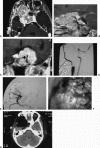
(A) A 27-year-old female with two previous resections and orbital exoneration and previous radiation therapy and chemotherapy for recurrent malignant meningioma. The patient presented with tumor regrowth and symptoms. Axial T1 MRI shows tumor within the cavernous sinus and orbital regions. (B) Sagittal T1 MRI with gadolinium showing the petrous and the cavernous ICA within the tumor. (C) Coronal T1 MRI with gadolinium showing ICA within the tumor. This finding prevented the previous surgeons from achieving complete resection. The patient failed BTO in our evaluation. (D) The patient underwent radial artery EC-IC bypass. Intraoperative angiogram above showing the radial artery graft. ICA has been occluded with a clip. (E) Angiogram showing flow into the MCA territory via the graft. (F) Intraoperative photograph showing tumor adherence to the wall of the cavernous ICA. Tumor resection was staged for the second day following EC-IC bypass and deliberate occlusion of the ICA proximal and distal to the tumor. (G) Postoperative axial CT scan showing tumor resection bed. MRI, magnetic resonance imaging; ICA, internal carotid artery; BTO, balloon occlusion testing; EC-IC, external carotid-internal carotid; MCA, middle carotid artery; CT, computed tomography.
Figure 7
Figure 7
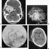
(A) A 45-year-old male with two previous resections of an atypical meningioma. The patient has received radiosurgery and chemotherapy. Previous resections were limited due to incorporation within the tumor of the superior division M2 (arrow). (B) Three-dimensional CT reconstruction of the blood vessels' relationship to the tumor. M2 division within the tumor shown (arrow). (C) Intraoperative angiogram shows radial artery EC-IC bypass to the distal portion of the involved M2 division. (D) Postoperative CT scan showing tumor resection within the sylvian fissure. CT, computed tomography; EC-IC, external carotid-internal carotid.

Similar articles

Cited by

References

    1. Yasargil M G, Krayenbuhl H A, Jacobson J H., 2nd Microneurosurgical arterial reconstruction. Surgery. 1970;67:221–233. - PubMed
    1. Lougheed W M, Marshall B M, Hunter M, Michel E R, Sandwith-Smyth H. Common carotid to intracranial internal carotid bypass venous graft. Technical note. J Neurosurg. 1971;34:114–118. - PubMed
    1. Sundt T M, Piepgras D G, Houser O W, et al. Interposition saphenous vein grafts for advanced occlusive disease and large aneurysms in the posterior circulation. J Neurosurg. 1982;56:205–215. - PubMed
    1. Spetzler R F, Fukushima T, Martin N, et al. Petrous carotid-to-intradural carotid saphenous vein graft for intracavernous giant aneurysm, tumor, and occlusive cerebrovascular disease. J Neurosurg. 1990;73:496–501. - PubMed
    1. Evans J J, Sekhar L N, Rak R, Stimac D. Bypass grafting and revascularization in the management of posterior circulation aneurysms. Neurosurgery. 2004;55:1036–1049. - PubMed