. 2005 Oct;3(10):e334.
doi: 10.1371/journal.pbio.0030334. Epub 2005 Sep 27.

Two-component signal transduction pathways regulating growth and cell cycle progression in a bacterium: a system-level analysis

Affiliations

Two-component signal transduction pathways regulating growth and cell cycle progression in a bacterium: a system-level analysis

Jeffrey M Skerker et al. PLoS Biol. 2005 Oct.

Abstract

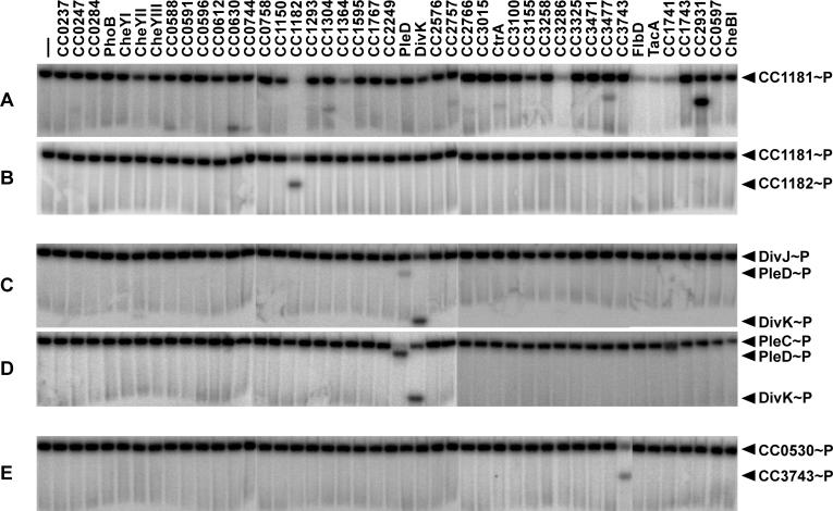
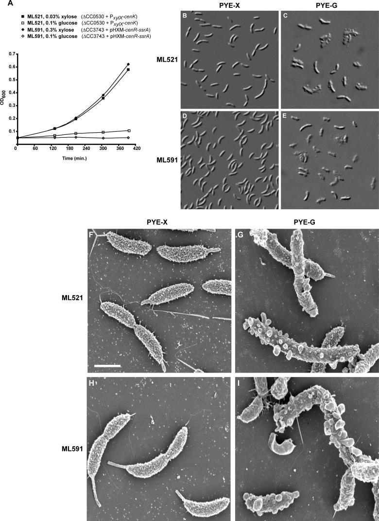
Two-component signal transduction systems, comprised of histidine kinases and their response regulator substrates, are the predominant means by which bacteria sense and respond to extracellular signals. These systems allow cells to adapt to prevailing conditions by modifying cellular physiology, including initiating programs of gene expression, catalyzing reactions, or modifying protein-protein interactions. These signaling pathways have also been demonstrated to play a role in coordinating bacterial cell cycle progression and development. Here we report a system-level investigation of two-component pathways in the model organism Caulobacter crescentus. First, by a comprehensive deletion analysis we show that at least 39 of the 106 two-component genes are required for cell cycle progression, growth, or morphogenesis. These include nine genes essential for growth or viability of the organism. We then use a systematic biochemical approach, called phosphotransfer profiling, to map the connectivity of histidine kinases and response regulators. Combining these genetic and biochemical approaches, we identify a new, highly conserved essential signaling pathway from the histidine kinase CenK to the response regulator CenR, which plays a critical role in controlling cell envelope biogenesis and structure. Depletion of either cenK or cenR leads to an unusual, severe blebbing of cell envelope material, whereas constitutive activation of the pathway compromises cell envelope integrity, resulting in cell lysis and death. We propose that the CenK-CenR pathway may be a suitable target for new antibiotic development, given previous successes in targeting the bacterial cell wall. Finally, the ability of our in vitro phosphotransfer profiling method to identify signaling pathways that operate in vivo takes advantage of an observation that histidine kinases are endowed with a global kinetic preference for their cognate response regulators. We propose that this system-wide selectivity insulates two-component pathways from one another, preventing unwanted cross-talk.


Figures

Figure 1
Figure 1. The C. crescentus Cell Cycle and Two-Component Signal Transduction
(A) Schematic diagram of progression through the C. crescentus cell cycle, as described in the text. The timing of key cell cycle and developmental events are indicated. Cell division is asymmetric, generating two distinct daughter cells. The stalked cell can immediately initiate DNA replication, whereas the swarmer cell must first differentiate into a stalked cell. (B) Diagram of a canonical two-component signal transduction system. On receipt of an input signal, the histidine kinase autophosphorylates on a conserved histidine residue. The phosphoryl group is then passed to the receiver domain of a cognate response regulator. Phosphorylation of the receiver domain typically activates the output domain, which can execute a variety of cellular tasks including initiating programs of gene expression, catalyzing metabolic reactions, or modifying protein–protein interactions.
Figure 2
Figure 2. Systematic Deletion of Two-Component Signal Transduction Genes
(A) Methodology used to generate chromosomal deletion strains. For each gene to be deleted, a suicide vector was constructed, with approximately 800-bp regions of homology upstream and downstream of the gene flanking a tetR cassette. See Materials and Methods and Figure S1 for details of plasmid construction. In a two-step process, deletion strains are isolated by selecting first for tetracycline resistance and then by sucrose counter-selection utilizing the sacB gene carried on the vector. Cells harboring the sacB gene die in the presence of sucrose. Hence, a deletion strain is identified as tetR/sucroseR. For nonessential genes, stable deletions are easily identified by screening 5–10 colonies after the two-step recombination. For essential genes, no tetR/sucroseR strains can be recovered (see text and Figure S1 for additional details). (B and C) Swarm plate analysis of 97 nonessential two-component deletion strains. (B) Map of strain positions in the swarm plates. Wild-type CB15N is in positions A1 and J10 for comparison to mutant strains. (C) PYE swarm plate after 3 d of growth at 30 °C. Swarm sizes and densities were scored visually and digital images analyzed in Matlab (MathWorks, Natick, Massachusetts, United States). Strains exhibiting swarm plate phenotypes are listed in Table 2, except for ΔCC1221 in position E1, which is deleted for a kinase erroneously annotated as a histidine kinase.
Figure 3
Figure 3. Morphology of Selected Deletion Strains
Deletion strains were harvested at mid-log phase and imaged using differential interference contrast microscopy. Strains: (A) wild-type CB15N, (B) ΔCC0138, (C) ΔCC0744, (D) ΔCC0909, (E) ΔCC1063, (F) ΔCC2482, (G) ΔCC3315, and (H) ΔCC3471.
Figure 4
Figure 4. Phosphotransfer Profiling Method
(A) Phosphotransfer profile experiments involve three separate reactions: (1) autophosphorylation of the histidine kinase (HK) by radiolabeled ATP, (2) phosphotransfer to a response regulator (RR), and (3) dephosphorylation of the response regulator. (B) Schematic of the phosphotransfer profiling technique. A single preparation of purified, autophosphorylated kinase (HK∼32P) is mixed with each response regulator from a given organism and analyzed for phosphotransfer by SDS-PAGE and autoradiography. The first lane shows a single band corresponding to the autophosphorylated histidine kinase and is used as a comparison for every other lane. Lanes 2–4 illustrate the three possible outcomes of a phosphotransfer reaction. In lane 2, phosphotransfer from HK to RR1 leads to the appearance of a band corresponding to RR1. In lane 3, phosphotransfer from HK to RR2 also occurs, but owing to high phosphatase activity (either autophosphatase or catalyzed by a bifunctional HK), the net result is production of inorganic phosphate (Pi) and the depletion of radiolabel from both the HK and RR2. In lane 4, no phosphotransfer occurs, and the lane is indistinguishable from lane 1. (C–H) Phosphotransfer profiling was performed for three E. coli kinases (EnvZ, CheA, and CpxA) against all 32 purified E. coli response regulators, with phosphotransfer incubation times of either 1 h (C, E, and G) or 10 s (D, F, and H). For these three histidine kinases, a comparison of the short and long time point profiles indicates a kinetic preference for only their in vivo cognate regulators: OmpR (C and D), CheY and CheB (E and F), and CpxR (G and H). After being examined for phosphotransfer, all gels are stained with Coomassie to verify equal loading of histidine kinase and response regulator in each lane (data not shown). For each kinase profiled, we purified only its soluble, cytoplasmic domain, either as a thioredoxin-His6 or a His6-MBP fusion, using standard metal affinity chromatography (see Materials and Methods). When necessary, we made successive N-terminal truncations until we identified a construct that produced active kinase in vitro, always preserving the H-box and ATP binding domain (details on constructs used are in Table S3). All response regulators were purified as full-length fusions to a thioredoxin-His6 tag. Purity was assessed by Coomassie staining, with each purified kinase domain and response regulator, except for E. coli FimZ, yielding an intense band of the correct approximate molecular weight (see Figure S5; Table S3).
Figure 5
Figure 5. Phosphotransfer Profiling of C. crescentus Histidine Kinases
Profiles for four purified C. crescentus kinases versus 44 purified response regulators were obtained by the method described for E. coli in Figure 4. (A) One-hour time point profile of the C. crescentus kinase CC1181. (B) Ten-second time point profile. Only CC1182, encoded in the same operon as CC1181 and the likely in vivo target, is phosphorylated at the short time point. Kinetic preference of C. crescentus histidine kinases for their cognate substrates was similarly demonstrated for five other operon pairs (data not shown). (C and D) Ten-second time point profiles of the orphan kinases DivJ and PleC, demonstrating phosphorylation of only their shared in vivo targets, PleD and DivK. (E) Phosphotransfer profiling of the previously uncharacterized essential orphan kinase CC0530 (CenK) reveals a single preferred substrate, CC3743 (CenR).
Figure 6
Figure 6. CC0530 (cenK) and CC3743 (cenR) Are Essential for Growth and Required for Cell Envelope Integrity
Growth curves for the ML521 (ΔCC0530 + PxylX-cenK) and ML591 (ΔCC3743 + pHXM-cenR-ssrA) depletion strains (A). Overnight cultures of each were grown in PYE plus xylose (PYE-X), washed with plain PYE, and diluted in PYE plus xylose or PYE plus glucose (PYE-G). After 12 h of growth in these conditions cells reached an optical density (OD600) level that could be measured (this time is plotted as “0 min”). Morphology was observed by light microscopy for the cenK depletion (ML521) after a total of 20 h in PYE plus xylose (B) or PYE plus glucose (C) and for the cenR depletion (ML591) after 20 h in PYE plus xylose (D) or PYE plus glucose (E). Scanning electron micrographs under identical conditions are shown for ML521 in PYE plus xylose (F) and PYE plus glucose (G) and for ML591 in PYE plus xylose (H) and PYE plus glucose (I). For (F–I), scale bar represents 1 μm. Depletion of either gene product led to an unusual, irregular blebbing of the cell surface. Cells were not motile, and had reduced stalk length.
Figure 7
Figure 7. Constitutive Activation of the CenK–CenR Pathway Leads to Dramatic Changes in Cell Morphology, Cell Lysis, and Death
Images are shown for strains grown overnight in PYE plus glucose and then diluted back to early log phase and grown for 5 h in PYE plus glucose (A, C, E, G, and I) or xylose (B, D, F, H, and J). In all panels, white arrows indicate cells with asymmetric bloating and black arrows indicate lysed cells. (A and B) ML603 (CB15N + pLXM-cenR + pJS71) expresses CenR alone from a low-copy vector. (C and D) ML675 (CB15N + pHXM-cenR) expresses CenR alone from a high-copy vector. (E and F) ML606 (CB15N + pLXM-cenR[D60E]) expresses, from a low-copy vector, a mutant of CenR that mimics constitutive phosphorylation. (G and H) ML607 (CB15N + pMR20 + pHXM-cenK cyto) expresses CenKcyto alone from a high-copy vector. (I and J) ML604 (CB15N + pLXM-cenR + pHXM-cenK cyto) expresses both CenR and CenKcyto, from low- and high-copy plasmids, respectively. (K) Growth curve for all strains from (A–J) grown in PYE supplemented with xylose [64,65].

References

    1. Ryan KR, Shapiro L. Temporal and spatial regulation in prokaryotic cell cycle progression and development. Annu Rev Biochem. 2003;72:367–394. - PubMed
    1. McAdams HH, Shapiro L. A bacterial cell-cycle regulatory network operating in time and space. Science. 2003;301:1874–1877. - PubMed
    1. Skerker JM, Laub MT. Cell-cycle progression and the generation of asymmetry in Caulobacter crescentus . Nat Rev Microbiol. 2004;2:325–337. - PubMed
    1. Iba H, Fukuda A, Okada Y. Rate of major protein synthesis during the cell cycle of Caulobacter crescentus . J Bacteriol. 1978;135:647–655. - PMC - PubMed
    1. Stock AM, Robinson VL, Goudreau PN. Two-component signal transduction. Annu Rev Biochem. 2000;69:183–215. - PubMed

Publication types

MeSH terms