. 2005 Dec;16(12):5514-27.
doi: 10.1091/mbc.e05-05-0410. Epub 2005 Sep 21.

Phosphorylation of N-cadherin-associated cortactin by Fer kinase regulates N-cadherin mobility and intercellular adhesion strength

Affiliations

Phosphorylation of N-cadherin-associated cortactin by Fer kinase regulates N-cadherin mobility and intercellular adhesion strength

Tarek Y El Sayegh et al. Mol Biol Cell. 2005 Dec.

Abstract

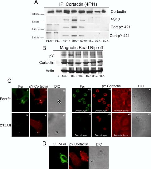
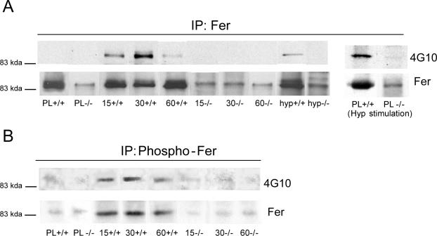
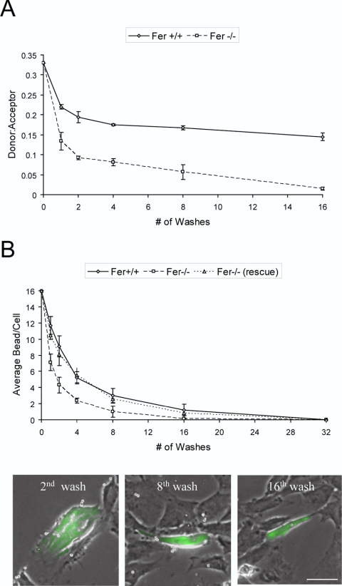
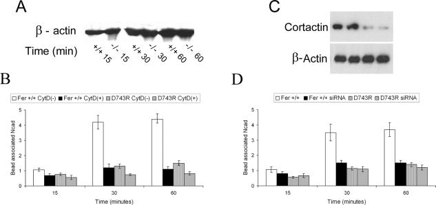
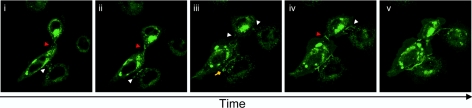
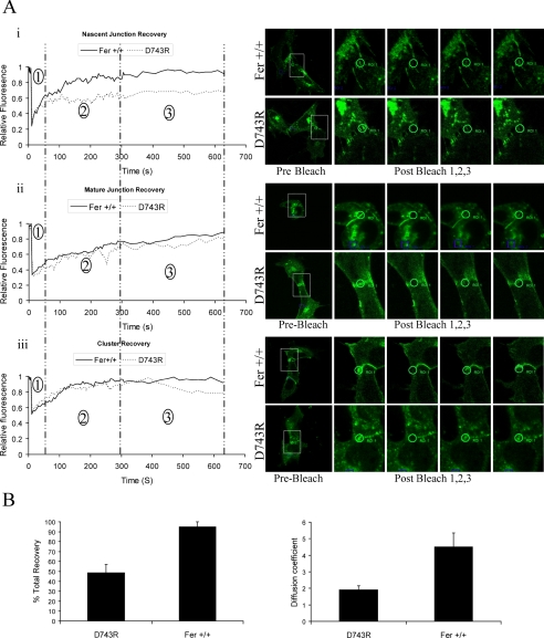
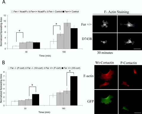
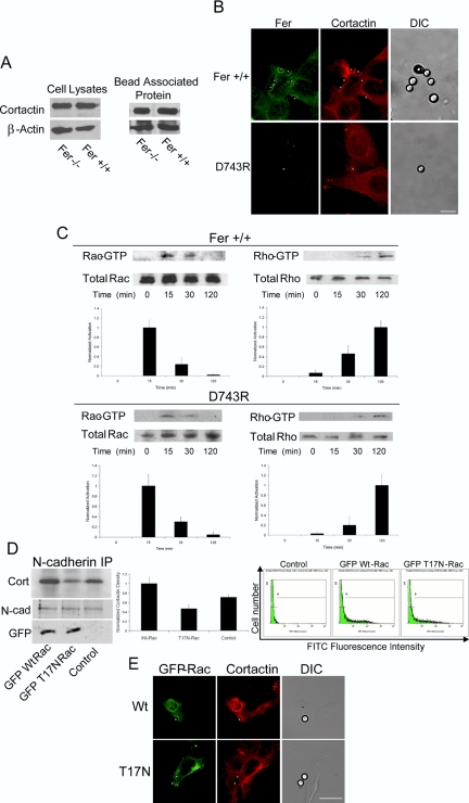
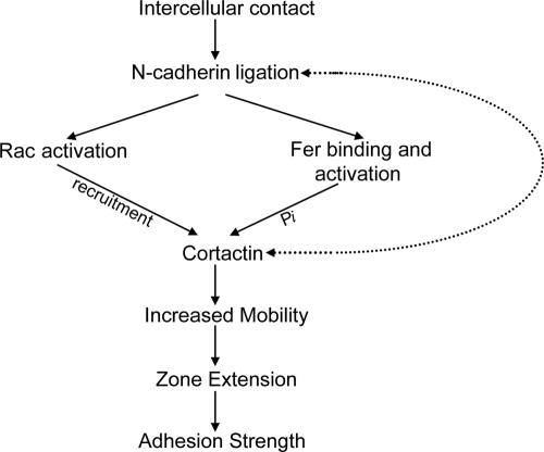
Cortactin regulates the strength of nascent N-cadherin-mediated intercellular adhesions through a tyrosine phosphorylation-dependent mechanism. Currently, the functional significance of cortactin phosphorylation and the kinases responsible for the regulation of adhesion strength are not defined. We show that the nonreceptor tyrosine kinase Fer phosphorylates cadherin-associated cortactin and that this process is involved in mediating intercellular adhesion strength. In wild-type fibroblasts N-cadherin ligation-induced transient phosphorylation of Fer, indicating that junction formation activates Fer kinase. Tyrosine phosphorylation of cortactin after N-cadherin ligation was strongly reduced in fibroblasts expressing only catalytically inactive Fer (D743R), compared with wild-type cells. In wild-type cells, N-cadherin-coated bead pull-off assays induced fourfold greater endogenous N-cadherin association than in D743R cells. Fluorescence recovery after photobleaching showed that GFP-N-cadherin mobility at nascent contacts was 50% faster in wild-type than D743R cells. In shear wash-off assays, nascent intercellular adhesion strength was twofold higher in wild-type than D743R cells. Cortactin recruitment to adhesions was independent of Fer kinase activity, but was impacted by N-cadherin ligation-provoked Rac activation. We conclude that N-cadherin ligation induces Rac-dependent cortactin recruitment and Fer-dependent cortactin phosphorylation, which in turn promotes enhanced mobilization and interaction of surface expressed N-cadherin in contacting cells.


Figures

Figure 1.
Figure 1.
Reduced cortactin tyrosine phosphorylation upon N-cadherin ligation in D743R fibroblasts. (A) Wild-type (+/+) or D743R (–/–) cells were allowed to attach to N-cad-Fc-coated bacteria-grade Petri-dishes or poly-l-lysine (PL)-treated tissue-culture dishes for 15, 30, or 60 min. Cell lysates were prepared and cortactin was immunoprecipitated (IP) and subjected to sequential immunoblotting analysis using antibodies to cortactin (SCBT H-191), phosphotyrosine (4G10), or phospho-cortactin recognizing pY421 (CortPY421) or pY486 (CortPY486). (B) Ncad-Fc-coated magnetic beads were applied to wild-type (+/+) or D743R (–/–) cells for 15, 30, or 60 min. N-cad-associated proteins were then isolated from cell lysates as described in Materials and Methods and assessed by immunoblotting with antibodies to phosphotyrosine (pY), cortactin or β-actin. (C) (i–vi) Ncad-Fc-coated beads or (vii–xiv) homotypic donor cells were applied to underlying wild-type (+/+) or D743R (–/–) cells for 15 min. Cells were fixed and subjected to immunofluorescence staining for Fer or pY421 cortactin, and the position of the beads or donor cells was determined by differential interference contrast (DIC) microscopy and or z-axis-oriented optical sectioning. Optical sections of donor cells (vii, viii, xi, and xii) or confluent underlying acceptor monolayers (ix and xiii). Results shown in i–vi are representative of ∼90% of bead-bound cells (42 and 39 of 45 examined beads bound wild-type and D743R cells, respectively), and (vii–xiv) 100% of observed bound donor cells {90 cells evaluated for each cell type). (D) Ncad-Fc-coated beads were applied for 15 min to D743R cells transfected with Fer-GFP. Fer-GFP was detected by direct fluorescence, pY421 was revealed by immunofluorescence and the position of the beads was revealed by DIC microscopy. These results are characteristic of ∼80% of the Fer-GFP-transfected cells (24 out of 30 examined positively transfected cells). Bars, 10 μm
Figure 2.
Figure 2.
Fer is tyrosine phosphorylated upon N-cadherin ligation. (A and B) Wild-type (+/+) or D743R (–/–) cells were attached to N-cad-Fc-coated Petri-dishes or poly-l-lysine (PL)-treated tissue-culture dishes for 15, 30, or 60 min and were subsequently treated with hyperosmotic media as indicated (hyp). Cell lysates were prepared and immunoprecipitated with antibodies to Fer or to phospho-Fer. Immunoprecipitates were immunoblotted with antibodies to phosphotyrosine (4G10) or Fer, as indicated.
Figure 3.
Figure 3.
Fer kinase activity is required for the development of adhesion strength in newly formed N-cadherin-mediated contacts. (A) Labeled donor wild-type (+/+) or D743R (–/–) cells were seeded onto confluent acceptor monolayers of the same genotype for 15 min, and adhesion strength was assessed by quantifying attached cells after washing as indicated. (B) Ncad-Fc-coated beads were applied onto monolayers of wild-type (+/+), D743R (–/–), or D743R cells transfected with Fer-GFP (rescue) for 15 min. Beads associated with cells were then quantified after washes as indicated by DIC microscopy or combined fluorescence and DIC microscopy in the case of the rescue cells. Bar, 20 μm.
Figure 4.
Figure 4.
Fer kinase influences actin-dependent cell surface organization of N-cadherin upon ligation. (A) Ncad-Fc-coated magnetic beads were applied to wild-type (+/+) or D743R (–/–) cells for 15, 30, or 60 min, and bead-associated actin was assessed as described in Materials and Methods. (B) The magnetic bead rip-off assay described above was repeated with cells treated with cytochalasin D or not, as indicated. Bead-associated N-cadherin was quantified and expressed relative to that seen in wild-type cells at 15 min in the absence of cytochalasin D. (C) Wild-type (+) or D743R (–) cells were treated with vehicle (V) or a specific RNAi against cortactin (S) as described in Materials and Methods. The cortactin content of whole cell lysates was assessed by Western blotting. (D) Magnetic bead rip-off assay in cortactin RNAi or vehicle control-treated samples. Bead-associated N-cadherin was quantified and expressed relative to that seen in wild-type cells at 15 min in vehicle controls.
Figure 5.
Figure 5.
N-cadherin-mediated intercellular contacts formation: Full-length N-cad-GFP-transfected Rat-2 cells in close proximity over an 8-h time course reveal the dynamic nature of contact formation and instability. Initial contact formation indicated by white arrows in i) and iii). Red arrow indicates a continuously remodeling unstable contact while white arrows (i, ii, and iv) indicate position of progressive contact formation. (i and iv) Radial pattern of N-cadherin at nascent contacts; ii) contact zone extension and stabilization (white arrow) and concurrent remodeling and loss of contact formation (red arrow); (iii) two zones of nascent contact formation (white arrows) and highly mobile peripheral cluster, which extends through contact zone (yellow diamond arrow); (iv) radial pattern representative of nascent contact formation (white arrow); (v) mobile cluster extends through contact zone. Red arrow indicates continued remodeling and junction instability.
Figure 6.
Figure 6.
Fer kinase activity influences N-cadherin mobility. (A) FRAP of N-cad-GFP in transfected wild-type and D743R cells for nascent (i) and mature (ii) junctions in 3.8-μm circular regions (shown in green on micrographs). Typical relative fluorescence plots (left) and corresponding micrographs (right) are presented for each experiment. Control (unbleached) regions in micrographs are in blue squares. (B) Quantification of FRAP analysis of wild-type and D743R cells bound to N-cad-Fc-coated substrata in zones of contact extension reveals significantly higher rate (p < 0.001) (right), and total recovery of fluorescence (left) in wild-type cells.
Figure 7.
Figure 7.
Fer kinase activity is required for efficient contact zone extension on an N-cadherin-based substrate. (A) Spreading of wild-type and D743R cells on N-cad-Fc or fibronectin-coated surfaces was quantified at 30 and 180 min as described in Materials and Methods (left). Images of F-actin-stained wild-type and D743 cells spreading on Ncad-Fc surfaces at 30 and 180 min are shown on right. (B) Spreading of wild-type and D743R cells depleted of endogenous cortactin by RNAi and concomitantly transfected with RNAi-resistant GFP-tagged cortactin constructs on N-cad-Fc-coated substrata as quantified at 30 and 180 min. Wild type (WT-cort) cortactin or a construct in which tyrosine residues 421, 466, and 482 were mutated to phenylalanines (F-cort) were used. Images of Fer+/+ cells at 180 min. *p < 0.05 between groups. Bars, 20 μm.
Figure 8.
Figure 8.
Cortactin recruitment to nascent N-cadherin ligation depends on transient Rac activation but is independent of Fer kinase activity. (A) Equivalent expression of cortactin in whole cell lysates of equal protein concentration in D743R and Fer+/+ fibroblasts. Ncad-Fc-coated magnetic bead pull-off after a 30-min incubation reveals equivalent amount of cortactin recruited to sites of N-cadherin ligation. (B) Immunofluorescence staining of Fer or cortactin in Ncad-Fc bead bound D743R and Fer+/+ cells after 15-min incubation reveal that recruitment of cortactin to sites of N-cadherin ligation is independent of Fer kinase activity. Results shown representative of ∼90% of observed bead bound cells {47 and 44 of 50 examined bead bound wild-type and D743R cells, respectively}. (C) N-cadherin engagement induces time-dependent activation of various rho GTPases. Fer+/+ fibroblasts bound to N-cad-Fc-coated 60-mm microbiological plastic dishes were harvested at distinct time points during the first 2 h of attachment. The GTP-bound forms of Rac and Rho were captured by GST-PBD and GST-Rhotekin, respectively. Results are representative of three independent experiments. (D) Recruitment of cortactin to areas of nascent N-cadherin ligation is dependent on locally activated Rac. Sorted Rat-2 cells transfected with wild-type and dominant negative GFP-tagged Rac constructs were attached to Ncad-Fc-coated substrata before lysis and N-cadherin immunoprecipitation. Blots probed for cortactin reveal reduced association of cortactin in T17N-transfected cells when compared with wild-type or controls (p < 0.05). Results are representative of three independent experiments. (E) Use of wild-type and negative (T17N) GFP-tagged Rac constructs demonstrates association of both constructs with areas of N-cadherin ligation and dependence of cortactin recruitment to areas of early N-cadherin ligation on Rac activation. Beads were incubated onto cells for 20 min. Results shown are consistent with 75% of bead bound cells {39 and 35 of 50 examined bead-bound Wt-Rac- and T17N-transfected cells, respectively}. Bar, 20 μm
Figure 9.
Figure 9.
Scheme outlining signaling events downstream of N-cadherin ligation that participate in subsequent enhancement of intercellular adhesion strength.

Similar articles

Cited by

References

    1. Adams, C. L., Chen, Y. T., Smith, S. J., and Nelson, W. J. (1998). Mechanisms of epithelial cell-cell adhesion and cell compaction revealed by high-resolution tracking of E-cadherin-green fluorescent protein. J. Cell Biol. 142, 1105–1119. - PMC - PubMed
    1. Adams, C. L., Nelson, W. J., and Smith, S. J. (1996). Quantitative analysis of cadherin-catenin-actin reorganization during development of cell-cell adhesion. J. Cell Biol. 135, 1899–1911. - PMC - PubMed
    1. Arora, P. D., Glogauer, M., Kapus, A., Kwiatkowski, D. J., and McCulloch, C. A. (2004). Gelsolin mediates collagen phagocytosis through a rac-dependent step. Mol. Biol. Cell 15, 588–599. - PMC - PubMed
    1. Arregui, C., Pathre, P., Lilien, J., and Balsamo, J. (2000). The nonreceptor tyrosine kinase fer mediates cross-talk between N-cadherin and beta1-integrins. J. Cell Biol. 149, 1263–1274. - PMC - PubMed
    1. Ben-Dor, I., Bern, O., Tennenbaum, T., and Nir, U. (1999). Cell cycle-dependent nuclear accumulation of the p94fer tyrosine kinase is regulated by its NH2 terminus and is affected by kinase domain integrity and ATP binding. Cell Growth Differ. 10, 113–129. - PubMed

Publication types