. 2005 Dec;16(12):5493-501.
doi: 10.1091/mbc.e05-03-0268. Epub 2005 Sep 21.

PERK and GCN2 contribute to eIF2alpha phosphorylation and cell cycle arrest after activation of the unfolded protein response pathway

Affiliations

PERK and GCN2 contribute to eIF2alpha phosphorylation and cell cycle arrest after activation of the unfolded protein response pathway

Robert B Hamanaka et al. Mol Biol Cell. 2005 Dec.

Abstract

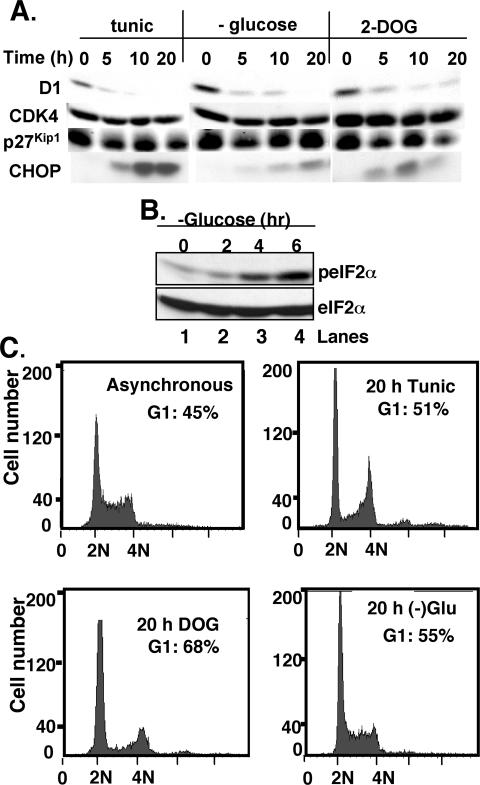
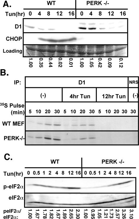
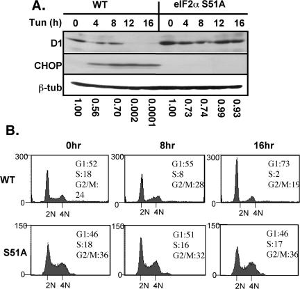
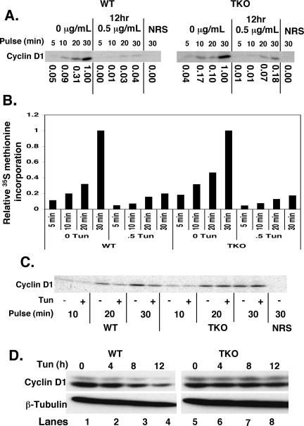
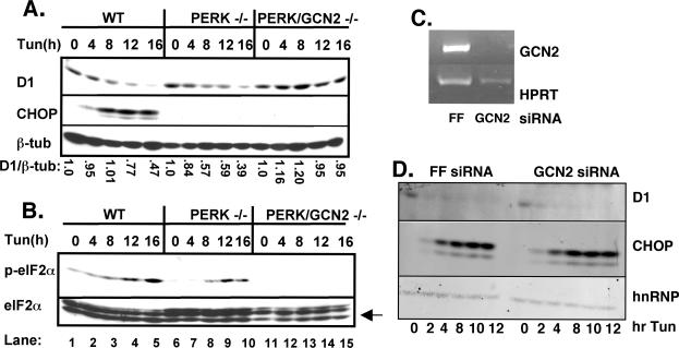
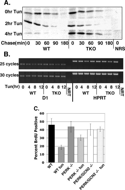
Exposure of cells to endoplasmic reticulum (ER) stress leads to activation of PKR-like ER kinase (PERK), eukaryotic translation initiation factor 2alpha (eIF2alpha) phosphorylation, repression of cyclin D1 translation, and subsequent cell cycle arrest in G1 phase. However, whether PERK is solely responsible for regulating cyclin D1 accumulation after unfolded protein response pathway (UPR) activation has not been assessed. Herein, we demonstrate that repression of cyclin D1 translation after UPR activation occurs independently of PERK, but it remains dependent on eIF2alpha phosphorylation. Although phosphorylation of eIF2alpha in PERK-/- fibroblasts is attenuated in comparison with wild-type fibroblasts, it is not eliminated. The residual eIF2alpha phosphorylation correlates with the kinetics of cyclin D1 loss, suggesting that another eIF2alpha kinase functions in the absence of PERK. In cells harboring targeted deletion of both PERK and GCN2, cyclin D1 loss is attenuated, suggesting GCN2 functions as the redundant kinase. Consistent with these results, cyclin D1 translation is also stabilized in cells expressing a nonphosphorylatable allele of eIF2alpha; in contrast, repression of global protein translation still occurs in these cells, highlighting a high degree of specificity in transcripts targeted for translation inhibition by phosphorylated eIF2alpha. Our results demonstrate that PERK and GCN2 function to cooperatively regulate eIF2alpha phosphorylation and cyclin D1 translation after UPR activation.


Figures

Figure 1.
Figure 1.
Glucose restriction triggers UPR activation and cyclin D1 loss. (A) Cell lysates prepared from asynchronously proliferating NIH-3T3 cells treated with 0.5 μg/ml tunicamycin (lanes 1–4), glucose free-media (lanes 5–8), or 10 mM 2-deoxyglucose (lanes 9–12) were subjected to immunoblot analysis with antibodies directed toward cyclin D1 (top), CDK4 (second panel), p27Kip1 (third panel), or CHOP (bottom). (B) Lysates prepared from cells cultured in glucose-free media for the indicated intervals were subjected to Western analysis with antibodies directed toward either phosphorylated (top) or total eIF2α. (C) Untreated NIH-3T3 cells or those cultured in the presence of tunicamycin, 2-DOG, or glucose-free media for 20 h were fixed and stained with propidium iodide and analyzed by flow cytometry. The percentage of cells with a 2N DNA content is presented in the inset.
Figure 2.
Figure 2.
PERK is not essential for repression of cyclin D1 translation during UPR activation. (A) Wild-type or PERK–/– fibroblasts were treated with 0.5 μg/ml tunicamycin for the indicated intervals. Equivalent amounts of total protein were subjected to Western blot analysis with antibodies specific for cyclin D1 or CHOP. (B) Lysates prepared from either wild-type or PERK–/– MEFs that had been pulsed for the indicated intervals with [35S]methionine after treatment with 0.5 μg/ml tunicamycin for 0, 4, or 12 h were subject to precipitation with normal rabbit antiserum (NRS) or a cyclin D1-specific mAb. Precipitated proteins were resolved by SDS-PAGE and visualized by autoradiography. (C) Lysates prepared as described in A were subjected to immunoblot analysis using antibodies directed toward serine 51 phosphorylated eIF2α or total eIF2α. The ratio of phosphorylated eIF2α relative to total eIF2α was determined by densitometry and was set to 1 in untreated cells within each cell type.
Figure 3.
Figure 3.
Translational inhibition of cyclin D1 and consequent cell cycle arrest are dependent upon eIF2α phosphorylation. (A) Wild-type or eIF2α S51A MEFs treated with 0.5 μg/ml tunicamycin for the indicated intervals were subject to Western blot analysis with antibodies specific for cyclin D1, CHOP, or β-tubulin. (B) FACS analysis of wild-type or eIF2α S51A MEFs treated with 0.5 μg/ml tunicamycin for the indicated intervals.
Figure 4.
Figure 4.
Continued cyclin D1 protein synthesis after treatment of cells lacking PERK/GCN2/PKR with tunicamycin. (A) PERK/GCN2/PKR triple knockout or wild-type fibroblasts were treated with the 0.5 μg/ml tunicamycin before pulse label with [35S]methionine. Cyclin D1 synthesis was assessed after metabolic labeling by precipitation with a cyclin D1-specific mAb or normal rabbit antiserum (NRS). (B) Total protein synthesis was monitored by precipitation of equivalent concentrations of total cellular lysates with TCA. Precipitated proteins were spotted onto filter paper, and new protein was quantified by scintillation counting. (C) TKO or wild-type fibroblasts were treated with 0.5 μg/ml tunicamycin for 6 h before a pulse with [35S]methionine for the indicated intervals. Protein concentrations were normalized to TCA-precipitable counts, and equivalent counts for each pulse length were immunoprecipitated for cyclin D1. (D) TKO or wild-type fibroblasts were treated with 0.5 μg/ml tunicamycin. Cyclin D1 and β-tubulin levels were assessed by Western analysis after separation of cell lysates on a denaturing polyacrylamide gel.
Figure 5.
Figure 5.
GCN2 cooperates with PERK in the inhibition of cyclin D1 translation after ER stress. Whole cell lysates prepared from wild-type, PERK–/–, or PERK/GCN2–/– cells treated with 0.5 μg/ml tunicamycin for the indicated intervals were resolved on a denaturing polyacrylamide gel and subject to Western blot analysis with antibodies specific for cyclin D1, CHOP, or β-tubulin (A) and phospho-eIF2α, total eIF2α, or CHOP (B). (C and D) Wild-type fibroblasts were infected with short hairpin vectors targeting GCN2. Forty-eight hours postinfection, cells were treated with 2 μg/ml tunicamycin for the indicated intervals and levels of cyclin D1, CHOP, and hnRNPK were determined by Western analysis, and knockdown of GCN2 was assessed by RT-PCR.
Figure 6.
Figure 6.
UPR activation does not affect cyclin D1 stability or transcription. (A) TKO or wild-type fibroblasts were treated with tunicamycin for 0, 2, or 4 h before a 30-min pulse with [35S]methionine. Cells were “chased” in media containing excess cold methionine for the indicated intervals. Cyclin D1 was precipitated with a D1 mAb and visualized by autoradiography. (B) TKO or wild-type fibroblasts were treated with 0.5 μg/ml tunicamycin for the indicated intervals. Total RNA was purified, reverse transcribed, and messages specific for cyclin D1 or HPRT were amplified by PCR. NRT indicates no RT input control. (C) Wild-type, PERK–/– and PERK/GCN2–/– fibroblasts proliferating on glass coverslips were left untreated or treated with tunicamycin (0.5 μg/ml) for 16 h. During the last 1.5 h, cells were pulsed with BrdU; BrdU incorporation was determined by immunofluorescence. Bars represent SD determined from three independent experiments.

Similar articles

Cited by

References

    1. Brewer, J. W., and Diehl, J. A. (2000). PERK mediates cell-cycle exit during the mammalian unfolded protein response. Proc. Natl. Acad. Sci. USA 97, 12625–12630. - PMC - PubMed
    1. Brewer, J. W., Hendershot, L. M., Sherr, C. J., and Diehl, J. A. (1999). Mammalian unfolded protein response inhibits cyclin D1 translation and cell-cycle progression. Proc. Natl. Acad. Sci. USA 96, 8505–8510. - PMC - PubMed
    1. Cherkasova, V. A., and Hinnebusch, A. G. (2003). Translational control by TOR and TAP42 through dephosphorylation of eIF2alpha kinase GCN2. Genes Dev. 17, 859–872. - PMC - PubMed
    1. Cox, J. S., Shamu, C. E., and Walter, P. (1993). Transcriptional induction of genes encoding endoplasmic reticulum resident proteins requires a transmembrane protein kinase. Cell 73, 1197–1206. - PubMed
    1. Cullinan, S. B., and Diehl, J. A. (2004). PERK-dependent activation of Nrf2 contributes to redox homeostasis and cell survival following ER stress. J. Biol. Chem. 279, 20076–20087. - PubMed

Publication types

MeSH terms