Spatial regulation of the cAMP-dependent protein kinase during chemotactic cell migration
- PMID: 16176981
- PMCID: PMC1242330
- DOI: 10.1073/pnas.0507072102
Spatial regulation of the cAMP-dependent protein kinase during chemotactic cell migration
Abstract
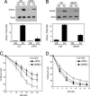
Historically, the cAMP-dependent protein kinase (PKA) has a paradoxical role in cell motility, having been shown to both facilitate and inhibit actin cytoskeletal dynamics and cell migration. In an effort to understand this dichotomy, we show here that PKA is regulated in subcellular space during cell migration. Immunofluorescence microscopy and biochemical enrichment of pseudopodia showed that type II regulatory subunits of PKA and PKA activity are enriched in protrusive cellular structures formed during chemotaxis. This enrichment correlates with increased phosphorylation of key cytoskeletal substrates for PKA, including the vasodilator-stimulated phosphoprotein (VASP) and the protein tyrosine phosphatase containing a PEST motif. Importantly, inhibition of PKA activity or its ability to interact with A kinase anchoring proteins inhibited the activity of the Rac GTPase within pseudopodia. This effect correlated with both decreased guanine nucleotide exchange factor activity and increased GTPase activating protein activity. Finally, inhibition of PKA anchoring, like inhibition of total PKA activity, inhibited pseudopod formation and chemotactic cell migration. These data demonstrate that spatial regulation of PKA via anchoring is an important facet of normal chemotactic cell movement.
Figures


Similar articles
-
Regulation of VASP serine 157 phosphorylation in human neutrophils after stimulation by a chemoattractant.J Leukoc Biol. 2007 Nov;82(5):1311-21. doi: 10.1189/jlb.0206107. Epub 2007 Aug 7. J Leukoc Biol. 2007. PMID: 17684042
-
Promotion of PDGF-induced endothelial cell migration by phosphorylated VASP depends on PKA anchoring via AKAP.Mol Cell Biochem. 2010 Feb;335(1-2):1-11. doi: 10.1007/s11010-009-0234-y. Epub 2009 Aug 27. Mol Cell Biochem. 2010. PMID: 19711176
-
Regulation of brefeldin A-inhibited guanine nucleotide-exchange protein 1 (BIG1) and BIG2 activity via PKA and protein phosphatase 1gamma.Proc Natl Acad Sci U S A. 2007 Feb 27;104(9):3201-6. doi: 10.1073/pnas.0611696104. Epub 2007 Feb 21. Proc Natl Acad Sci U S A. 2007. PMID: 17360629 Free PMC article.
-
Regulation of actin-based cell migration by cAMP/PKA.Biochim Biophys Acta. 2004 Jul 5;1692(2-3):159-74. doi: 10.1016/j.bbamcr.2004.03.005. Biochim Biophys Acta. 2004. PMID: 15246685 Review.
-
A-kinase-anchoring proteins and cytoskeletal signalling events.Biochem Soc Trans. 2003 Feb;31(Pt 1):87-9. doi: 10.1042/bst0310087. Biochem Soc Trans. 2003. PMID: 12546660 Review.
Cited by
-
Endothelial CD99 signals through soluble adenylyl cyclase and PKA to regulate leukocyte transendothelial migration.J Exp Med. 2015 Jun 29;212(7):1021-41. doi: 10.1084/jem.20150354. Epub 2015 Jun 22. J Exp Med. 2015. PMID: 26101266 Free PMC article.
-
Abl-interactor-1 (Abi1) has a role in cardiovascular and placental development and is a binding partner of the alpha4 integrin.Proc Natl Acad Sci U S A. 2011 Jan 4;108(1):149-54. doi: 10.1073/pnas.1012316108. Epub 2010 Dec 20. Proc Natl Acad Sci U S A. 2011. PMID: 21173240 Free PMC article.
-
cAMP Bursts Control T Cell Directionality by Actomyosin Cytoskeleton Remodeling.Front Cell Dev Biol. 2021 May 20;9:633099. doi: 10.3389/fcell.2021.633099. eCollection 2021. Front Cell Dev Biol. 2021. PMID: 34095108 Free PMC article.
-
Anchoring of protein kinase A by ERM (ezrin-radixin-moesin) proteins is required for proper netrin signaling through DCC (deleted in colorectal cancer).J Biol Chem. 2015 Feb 27;290(9):5783-96. doi: 10.1074/jbc.M114.628644. Epub 2015 Jan 9. J Biol Chem. 2015. PMID: 25575591 Free PMC article.
-
Guanylate cyclase and cyclic GMP-dependent protein kinase regulate agrin signaling at the developing neuromuscular junction.Dev Biol. 2007 Jul 15;307(2):195-201. doi: 10.1016/j.ydbio.2007.04.021. Epub 2007 Apr 24. Dev Biol. 2007. PMID: 17560564 Free PMC article.
References
Publication types
MeSH terms
Substances
Grants and funding
LinkOut - more resources
Full Text Sources
Miscellaneous

