Comparative Study
. 2005 Sep 21;25(38):8794-806.
doi: 10.1523/JNEUROSCI.2663-05.2005.

Proprioceptor regulation of motor circuit activity by presynaptic inhibition of a modulatory projection neuron

Affiliations
Comparative Study

Proprioceptor regulation of motor circuit activity by presynaptic inhibition of a modulatory projection neuron

Mark P Beenhakker et al. J Neurosci. .

Abstract

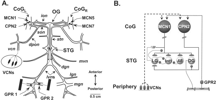
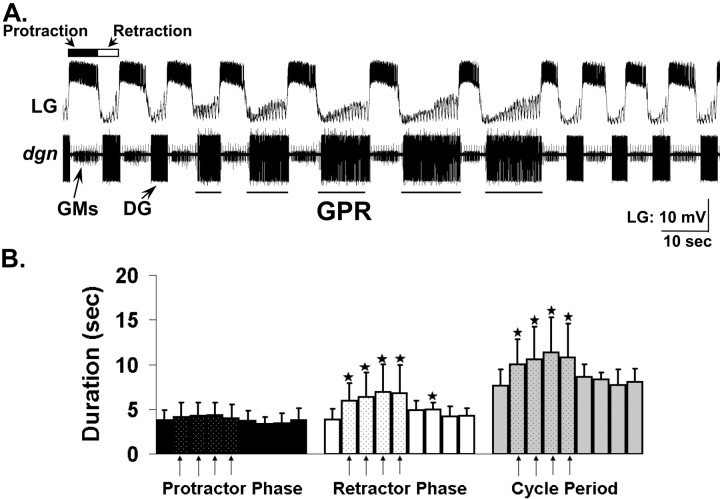
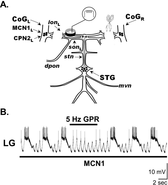
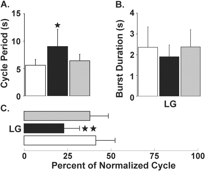
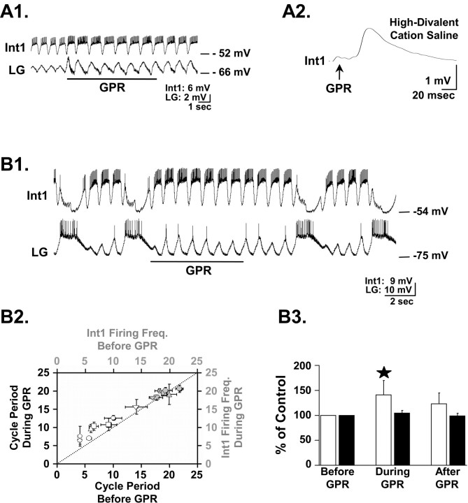
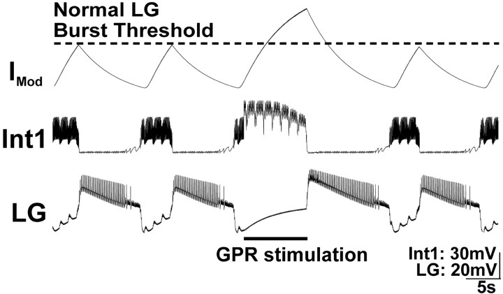
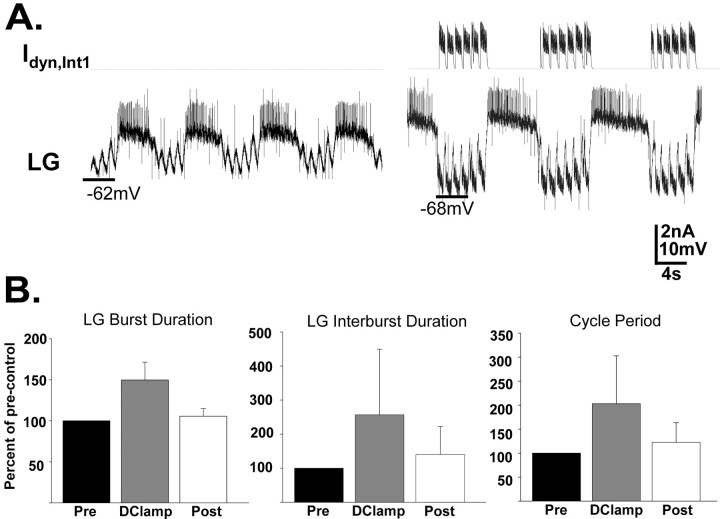
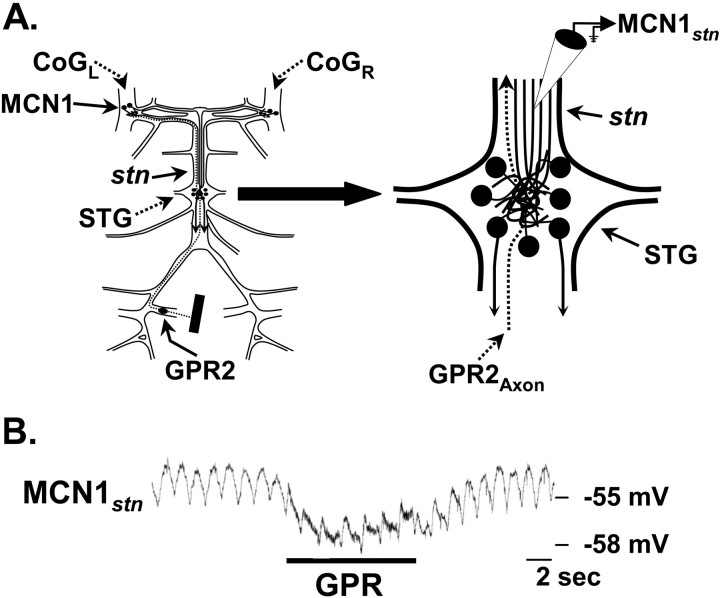
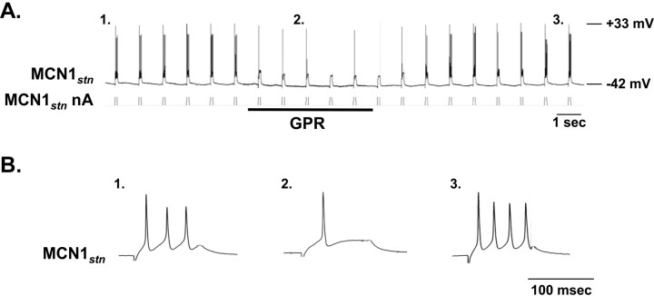
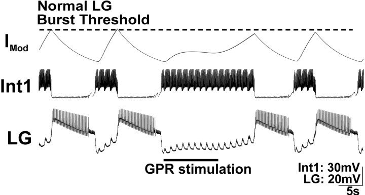
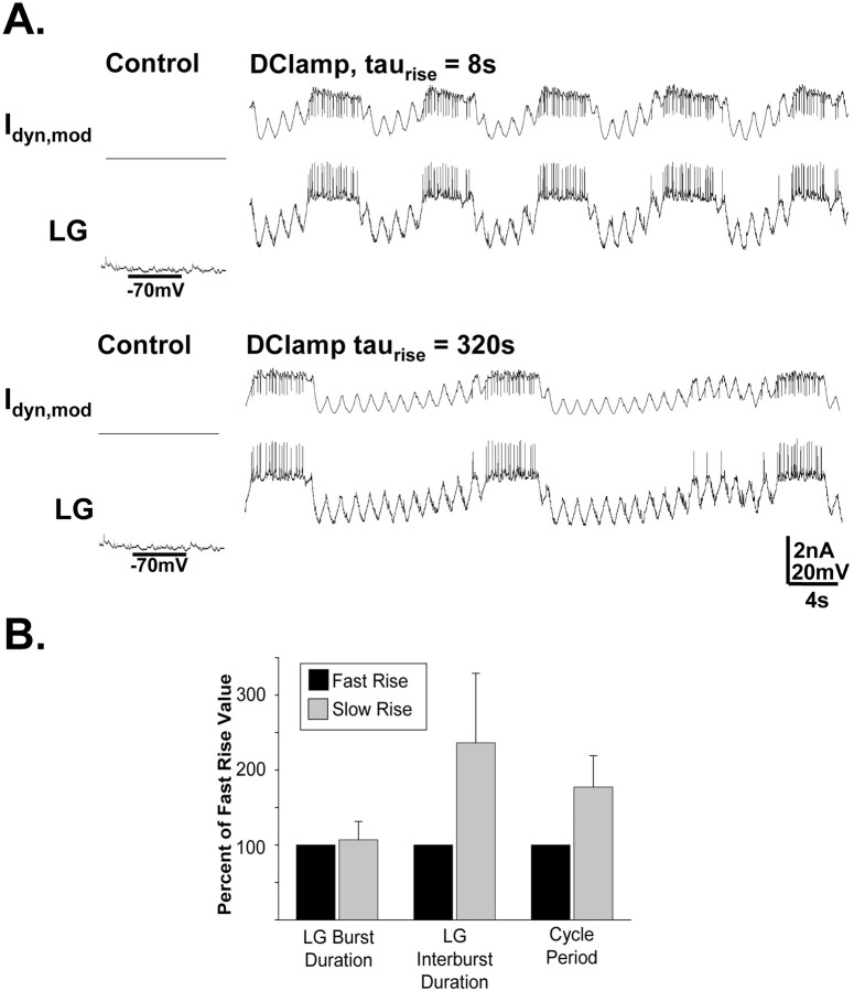
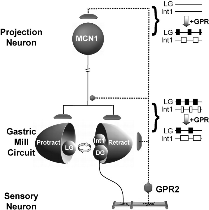
Phasically active sensory systems commonly influence rhythmic motor activity via synaptic actions on the relevant circuit and/or motor neurons. Using the crab stomatogastric nervous system (STNS), we identified a distinct synaptic action by which an identified proprioceptor, the gastropyloric muscle stretch receptor (GPR) neuron, regulates the gastric mill (chewing) motor rhythm. Previous work showed that rhythmically stimulating GPR in a gastric mill-like pattern, in the isolated STNS, elicits the gastric mill rhythm via its activation of two identified projection neurons, modulatory commissural neuron 1 (MCN1) and commissural projection neuron 2, in the commissural ganglia. Here, we determine how activation of GPR with a behaviorally appropriate pattern (active during each gastric mill retractor phase) influences an ongoing gastric mill rhythm via actions in the stomato gastric ganglion, where the gastric mill circuit is located. Stimulating GPR during each retractor phase selectively prolongs that phase and thereby slows the ongoing rhythm. This selective action on the retractor phase results from two distinct GPR actions. First, GPR presynaptically inhibits the axon terminals of MCN1, reducing MCN1 excitation of all gastric mill neurons. Second, GPR directly excites the retractor phase neurons. Because MCN1 transmitter release occurs during each retractor phase, these parallel GPR actions selectively reduce the buildup of excitatory drive to the protractor phase neurons, delaying each protractor burst. Thus, rhythmic proprioceptor feedback to a motor circuit can result from a global reduction in excitatory drive to that circuit, via presynaptic inhibition, coupled with a phase-specific excitatory input that prolongs the excited phase by delaying the onset of the subsequent phase.


Figures

Figure 1.
Figure 1.
Schematic of the isolated STNS and the pathways by which two identified sensory systems influence the gastric mill circuit. A, The STNS consists of the unpaired STG and oesophageal (OG) ganglia, the paired CoG, plus the connecting and peripheral nerves. In each CoG, there is a single copy of each identified CoG projection neuron, including MCN1, MCN5, MCN7, and CPN2. The STNS receives sensory information from the proprioceptor neurons GPR1 and GPR2 and the mechanoreceptor VCNs. B, GPRs and VCNs each elicit the gastric mill rhythm by activating MCN1 and CPN2, which in turn activate the gastric mill circuit. The gastric mill CPG neurons LG and Int1 have reciprocal inhibitory connections and are influenced by the pyloric pacemaker neuron AB. The DG neuron is a gastric mill retractor motor neuron that innervates the gm4 muscle. Contraction of gm4 stretches the GPR-innervated muscles, thereby activating GPRs (Katz and Harris-Warrick, 1989). Synapse symbols: t-bars, excitation; filled circles, inhibition; resistor, electrical coupling. Nerves: dgn, dorsal gastric nerve; dpon, dorsal posterior oesophageal nerve; ion, inferior oesophageal nerve; lgn, lateral gastric nerve; lvn, lateral ventricular nerve; mgn, medial gastric nerve; mvn, medial ventricular nerve; son, superior oesophageal nerve; stn, stomatogastric nerve; vcn, ventral cardiac nerve.
Figure 2.
Figure 2.
GPR stimulation prolongs the retractor phase of the VCN-elicited gastric mill rhythm. A, Stimulating GPR (5 Hz intraburst frequency) in a behaviorally relevant pattern during the VCN-elicited gastric mill rhythm in the isolated stomatogastric nervous system slows the rhythm by selectively prolonging the retractor phase. B, Relative to pre-GPR stimulation levels, during each cycle when GPR was stimulated (arrows), there was a significant increase in the duration of the gastric mill retractor phase and in the duration of the cycle period, but there was no change in the protractor phase duration (*p < 0.05; n = 8; one-way repeated-measures ANOVA, Student–Newman–Keuls test). Each bar represents a single gastric mill cycle, with one cycle shown pre-GPR, five cycles shown during GPR, and four cycles shown post-GPR stimulation. Note the rapid return to control levels after GPR stimulation was terminated. Error bars indicate SD.
Figure 3.
Figure 3.
Influence of GPR on the MCN1-elicited gastric mill rhythm. A, Preparation used to study the MCN1-elicited gastric mill rhythm. The CoGs were eliminated by transection of the ions and sons, and selective MCN1 stimulation was accomplished by extracellular stimulation of the ion to elicit the gastric mill rhythm (Bartos et al., 1999). B, Stimulating GPR during the retractor phase of the MCN1-elicited gastric mill rhythm selectively prolonged that retractor phase. MCN1 stimulation (black bar, 30 Hz tonic) began before the presented recording segment and persisted for the duration of the recording shown. Most hyperpolarized Vm: LG, –75 mV.
Figure 4.
Figure 4.
Quantification of the GPR actions on the MCN1-elicited gastric mill rhythm. The pre-GPR (white bars), during GPR (black bars), and post-GPR stimulation (gray bars) conditions each represent the mean of five consecutive gastric mill cycles. A, GPR stimulation during the MCN1-elicited gastric mill rhythm reversibly prolonged the gastric mill cycle period by ∼60% (*p < 0.05; n = 6). B, C, Stimulating GPR during the MCN1-elicited gastric mill rhythm did not alter the LG neuron burst duration (p > 0.05; n = 6) (B) but reduced the fraction of a gastric mill cycle during which LG was active (LG duty cycle) by nearly 20% (**p < 0.01; n = 6) (C). Error bars indicate SD.
Figure 5.
Figure 5.
GPR excites Int1 but does not alter Int1 activity during the gastric mill rhythm. A1, In the isolated STG with no ongoing gastric mill rhythm, GPR stimulation (5Hz) excited Int1 and produced a concomitant increase in the amplitude of the subthreshold, pyloric-timed oscillations in the LG neuron. A2, Single GPR stimuli evoked constant latency EPSPs in Int1 in high divalent cation saline.The EPSP represents the average of eight Int1 responses to GPR stimulation. The relatively long latency to EPSP onset results from the ∼2 cm distance traveled by the GPR-elicited action potentials to reach the STG. B1, GPR stimulation (5 Hz) during an ongoing MCN1-elicited gastric mill rhythm prolonged the retractor phase without an evident change in Int1 activity. B2, GPR failed to enhance Int1 activity during ongoing gastric mill rhythms at times when it did prolong the gastric mill cycle period. The mean ± SD gastric mill cycle period (white symbols) and corresponding Int1 firing frequency (gray symbols) before and during GPR stimulation for eight preparations are shown, organized by symbol shape. The dotted line represents no change between the pre-GPR and during GPR conditions. B3, Int1 firing frequency (black bars) and gastric mill cycle period (white bars) data are normalized to precontrol conditions. In the same preparations, GPR stimulation increased the gastric mill cycle period (40.8 ± 28.9%; *p < 0.001; n = 8) but failed to alter Int1 firing frequency (4.5 ± 5.4%; p > 0.05; n = 8). Error bars indicate SD.
Figure 6.
Figure 6.
Increasing the strength of Int1 inhibition of the LG neuron during GPR stimulation prolongs both phases of the MCN1-elicited gastric mill rhythm in a computational model. GPR was stimulated during a retractor phase, as occurs in the biological system. In this version of the model, the strength of Int1 inhibition of LG was explicitly enhanced by GPR stimulation. The result was an increase in the duration of that retractor phase (Int1 active) as well as an increase in the subsequent protractor phase (LG active). Note that the rhythm returned to control levels in the next cycle. The modulatory current (IMod) trace represents the cycle-by-cycle buildup and decay of MCN1 excitation of the LG neuron. When GPR is silent, the LG neuron burst initiates when the level of modulatory current attains the level designated as “normal LG burst threshold.” Most hyperpolarized Vm: Int1, –65 mV; LG, –55 mV.
Figure 7.
Figure 7.
Increasing the strength of Int1 inhibition of the LG neuron with the dynamic clamp in the biological preparation slows the gastric mill rhythm by prolonging both phases of the rhythm. A, During an ongoing MCN1-elicited gastric mill rhythm, the dynamic clamp was used to increase the strength of Int1 inhibition of the LG neuron (Idyn,Int1). This increased inhibition led to a prolonged retractor phase, as well as a longer duration of each subsequent protractor phase. The vertical lines occurring periodically during the LG burst (rising above and below the action potentials) and interburst represent artifacts that occur when recording in discontinuous current-clamp mode while performing nerve (ion) stimulation. B, Quantitative analysis supporting the result represented in A. Under these conditions, GPR stimulation reversibly prolonged the LG burst duration, LG interburst duration, and gastric mill cycle period (p < 0.01; n = 6). Error bars indicate SD.
Figure 8.
Figure 8.
GPR stimulation hyperpolarizes the STG terminals of MCN1. A, Schematic of the isolated STNS (left) with an expanded view of the STG (right) to indicate the location of the intra-axonal recording site for MCN1stn. B, Intracellular recording of MCN1stn revealed a hyperpolarizing response to GPR stimulation (5 Hz). Note the subthreshold pyloric-timed oscillations in MCN1stn. The thickened MCN1stn trace during GPR stimulation represents the stimulus artifact.
Figure 9.
Figure 9.
GPR stimulation inhibits MCN1 action potential initiation within the STG neuropil. A, Injecting depolarizing current into MCN1stn initiates action potentials within the STG neuropil (Coleman and Nusbaum, 1994). Constant duration (100 ms) and amplitude (+2 nA) depolarizing pulses injected into MCN1stn produced a regular number (3–4) of action potentials per pulse. This number was reversibly reduced during GPR stimulation (5 Hz). Note the delay of several seconds after GPR stimulation before the number of MCN1 action potentials per pulse returns to control levels. B, Expanded traces from A of single pulses before (1), during (2), and after (3) GPR stimulation.
Figure 10.
Figure 10.
The GPR action on the MCN1-elicited gastric mill rhythm is best mimicked by GPR inhibition of MCN1STG in a computational model of the MCN1-elicited gastric mill rhythm. In this version of the model, GPR stimulation presynaptically inhibited MCN1STG, thereby reducing MCN1 actions onto the LG neuron. As a result, the amplitude of the modulatory current (IMod) induced in the LG neuron by MCN1 is reduced during GPR stimulation. This effect persists after GPR stimulation because of the slow time constant of the GPR action. The GPR inhibition of MCN1STG slowed the LG escape from Int1 inhibition, prolonging the retractor phase. Note that, in contrast, the protractor phase duration was not altered. Most hyperpolarized Vm: Int1, –65 mV; LG, –55 mV.
Figure 11.
Figure 11.
The rate of rise of MCN1-like modulation of the LG neuron selectively regulates the retractor phase duration. A, In this experiment, in place of MCN1 stimulation, the biological LG neuron was injected with a dynamic clamp version of the modulatory current (Idyn,mod) that likely represents the current provided by MCN1 (Swensen and Marder, 2000) (see Materials and Methods). The only parameter that differed during the dynamic clamp current injection in the two traces shown was the rate of rise of the current amplitude during each LG interburst. Note that the slower rate of rise (bottom trace) caused a slower rhythm, which resulted primarily from a selectively prolonged LG interburst (retractor-like phase). Both LG traces are from the same preparation. B, Injecting the dynamic clamp version of the modulatory current into LG, with a relatively long time constant for its rise in amplitude, consistently elicited gastric mill-like activity in LG that was slower (because of a selective increase in LG interburst duration) than that resulting from injection of the same current with a briefer time constant (p < 0.005; n = 5). Error bars indicate SD.
Figure 12.
Figure 12.
Working model of the GPR actions on the gastric mill system. Gastric mill-like rhythmic GPR stimulation can elicit the gastric mill rhythm (Blitz et al., 2004) by exciting the projection neuron MCN1 (and CPN2) in the commissural ganglia. During the gastric mill rhythm elicited by MCN1 (either directly or via activation of the VCN mechanosensory neurons), stimulating GPR in a behaviorally appropriate manner (during each retractor DG neuron burst) slows the gastric mill rhythm by selectively prolonging the retractor phase. This latter GPR action results from GPR inhibition of the axon terminals of MCN1 in the stomatogastric ganglion, in concert with its excitation of the retractor neurons Int1 and DG. Synapse symbols: t-bars, excitation; filled circles, inhibition. Activity symbols: filled boxes, LG action potential bursts; open boxes, Int1 action potential bursts.

Similar articles

Cited by

References

    1. Bartos M, Nusbaum MP (1997) Intercircuit control of motor pattern modulation by presynaptic inhibition. J Neurosci 17: 2247–2256. - PMC - PubMed
    1. Bartos M, Manor Y, Nadim F, Marder E, Nusbaum MP (1999) Coordination of fast and slow rhythmic neuronal circuits. J Neurosci 19: 6650–6660. - PMC - PubMed
    1. Beenhakker MP (2004) Sensory regulation of rhythmically active neuronal networks. PhD thesis, University of Pennsylvania.
    1. Beenhakker MP, Nusbaum MP (2003) Sensory regulation of a modulatory projection neuron at two separate locations. Soc Neurosci Abstr 29: 604.11.
    1. Beenhakker MP, Nusbaum MP (2004) Mechanosensory activation of a motor circuit by coactivation of two projection neurons. J Neurosci 24: 6741–6750. - PMC - PubMed

Publication types