. 2005 Oct 1;175(7):4265-73.
doi: 10.4049/jimmunol.175.7.4265.

Dendritic cell-specific intercellular adhesion molecule 3-grabbing nonintegrin/CD209 is abundant on macrophages in the normal human lymph node and is not required for dendritic cell stimulation of the mixed leukocyte reaction

Affiliations

Dendritic cell-specific intercellular adhesion molecule 3-grabbing nonintegrin/CD209 is abundant on macrophages in the normal human lymph node and is not required for dendritic cell stimulation of the mixed leukocyte reaction

Angela Granelli-Piperno et al. J Immunol. .

Abstract

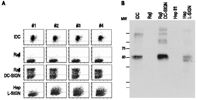
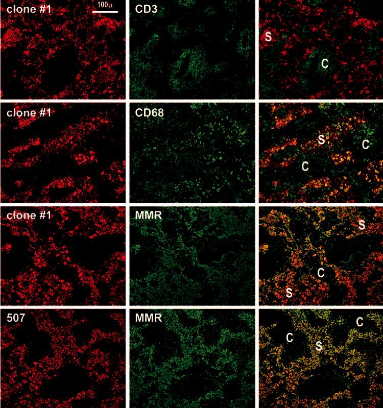
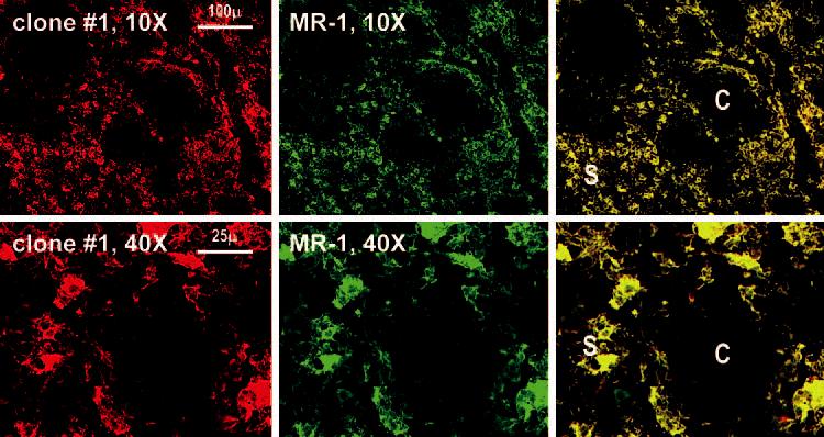
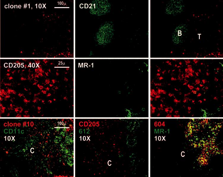
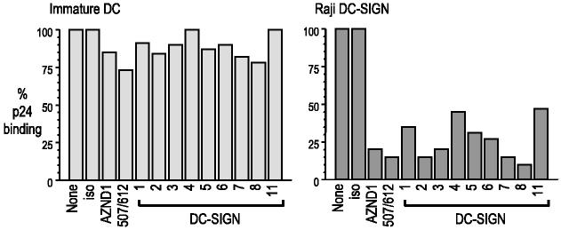
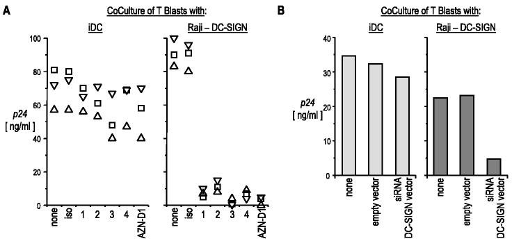
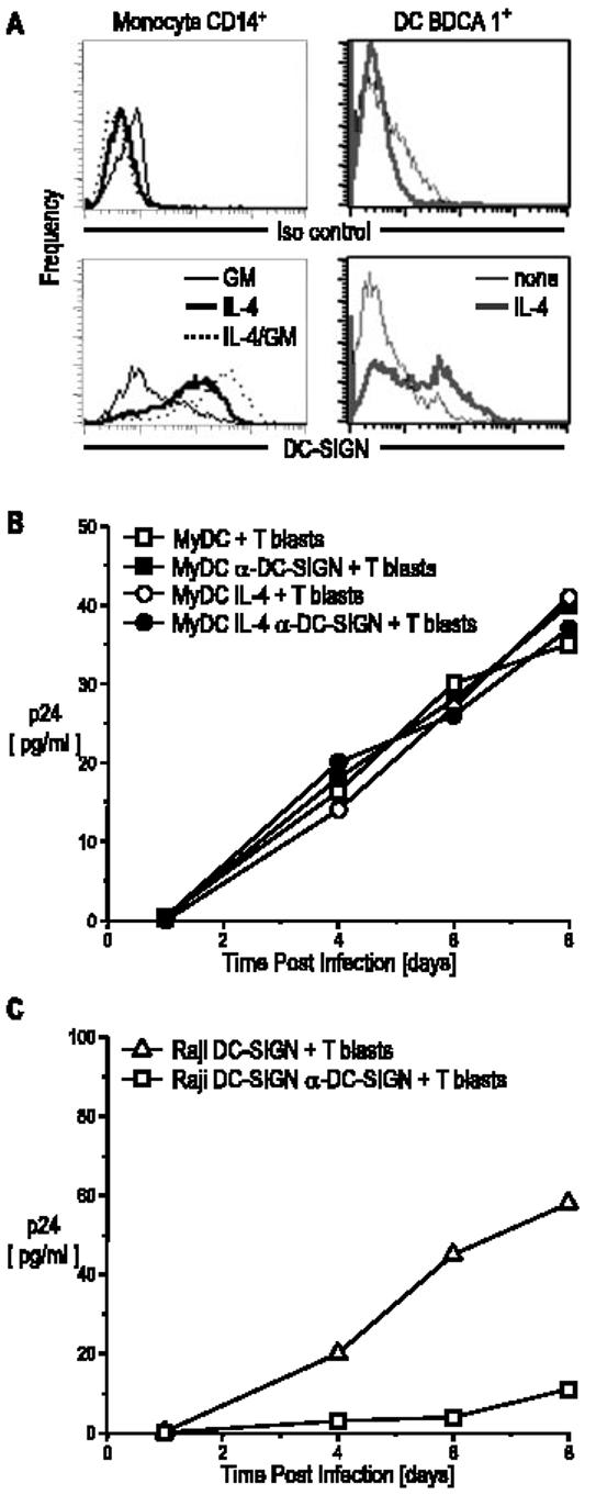
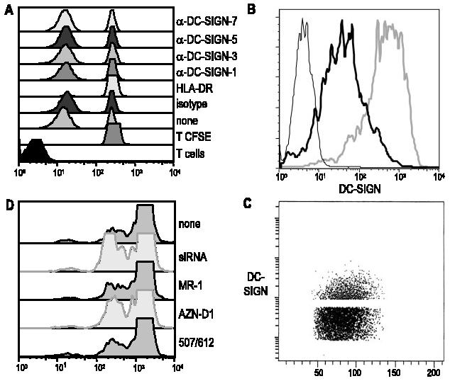
The C-type lectin dendritic cell-specific ICAM 3-grabbing nonintegrin (DC-SIGN)/CD209 efficiently binds several pathogens, including HIV-1. DC-SIGN is expressed on monocyte-derived DCs in culture, and importantly, it is able to sequester HIV-1 within cells and facilitate transmission of virus to CD4+ T cells. To investigate DC-SIGN function, we have generated new mAbs. We report in this study that these and prior anti-DC-SIGN mAbs primarily label macrophages in the medullary sinuses of noninflamed human lymph node. In contrast, expression is not detected on most DCs in the T cell area, except for occasional cells. We also noted that IL-4 alone can induce expression of DC-SIGN in CD14+ monocytes and circulating blood DCs. However, blockade of DC-SIGN with Abs and DC-SIGN small interfering RNA did not result in a major reduction in the capacity of these DCs to transfer HIV to T cells, confirming significant DC-SIGN-independent mechanisms. The blocking approaches did reduce HIV-1 transmission by DC-SIGN-transfected cells by >90%. DC-SIGN blockade also did not reduce the ability of DCs to stimulate T cell proliferation in the MLR. These results indicate that DC-SIGN has the potential to contribute to macrophage function in normal human lymph node, and that DCs do not require DC-SIGN to transmit HIV or to initiate T cell responses.


Figures

FIGURE 1
FIGURE 1
Reactivity of mAbs with DC-SIGN in transfected cells and DCs. A, Reactivity of anti-DC-SIGN mAb with DCs and transfectants expressing full-length DC-SIGN or L-SIGN. B, Reactivity by immunoblot of anti-DC-SIGN mAb 2. The indicated cells were lysates in RIPA buffer, equivalent to 3 × 10⁵ cells. After separation on SDS-PAGE, the lysates were blotted with anti-DC-SIGN mAb 2.
FIGURE 2
FIGURE 2
DC-SIGN staining is restricted to macrophages in the medullary sinuses (S) of lymph nodes. Two-color staining is shown, with red being the anti-DC-SIGN reagent, and green the other markers. First row, Clone 1 anti-DC-SIGN stains macrophages in the sinuses of the medulla and anti-CD3 stains T cells in the medullary cords (C). Second row, Clone 1 and anti-CD68 colabel medullary macrophages. Third row, Clone 1 and anti-macrophage mannose receptor (MMR) colabel macrophages in the lymph node medulla. Fourth row, Double staining using a commercial anti-DC-SIGN 507 and anti-mannose receptor mAbs.
FIGURE 3
FIGURE 3
Identical staining by clone 1 anti-DC-SIGN and another established anti-DC-SIGN mAb. Lymph node medulla sections were double stained with our clone 1 and the anti-DC-SIGN mAb MR-1.
FIGURE 4
FIGURE 4
Immunostaining of the lymph node cortex with anti-DC-SIGN mAbs. First row, The lymph node cortex is shown at ×10 after staining with anti-CD21 (green) to define the B cell follicles (B) and the unstained T cell region (T). Second row, Images at higher magnification within the T cell region reveal that most DCs defined by staining with anti-CD205 are DC-SIGN negative. Third row, The left panel is a double staining with our clone 10 (specific for DC-SIGN and L-SIGN) and CD11c; the middle panel is a double staining with clone 612 (specific for DC SIGN and L-SIGN) and DEC205; the right panel shows a double staining using anti-L-SIGN specific mAb (604) with MR-1.
FIGURE 5
FIGURE 5
Immunostaining of the lymph node cortex. Higher magnification of the cortex where rare fields can be found in which red DC-SIGN-positive cells colabel with CD11c and anti-CD205.
FIGURE 6
FIGURE 6
HIV-1 binding to DC-SIGN-expressing cells. Cells were incubated with anti-DC-SIGN mAbs or isotype control at 20 μg/ml for 20 min at room temperature and then infected with HIV-1 for 2 h at 37°C. Cells were washed and lysed in 0.5% Triton. The amount of p24 in the cell lysates was quantified by ELISA.
FIGURE 7
FIGURE 7
Effect of anti-DC-SIGN mAbs on HIV-1 transmission to T blasts. A, The indicated cells (left panel, immature DCs; right panel, Raji-DC-SIGN) were incubated with anti-DC-SIGN mAbs as described in Fig. 6, and then infected with HIV-1 BaL in the continued presence of mAb. The cells were washed four times, and 5 × 10⁴ infected cells were cocultured with 105 T blasts. After 5 days, p24 secreted in the culture supernatants was quantified by ELISA. Each symbol corresponds to an individual experiment. B, DC-SIGN-expressing cells were transduced with siRNA lentiviral vector or empty vector at an MOI of 20, sorted for low DC-SIGN expression, and infected with HIV-1 before addition to T cells. The resulting cells were cocultured and assayed as described above.
FIGURE 8
FIGURE 8
DC-SIGN expression is induced by IL-4 and is not required for HIV-1 transmission by blood myeloid DCs. A, CD14+ cells and BDCA1+ cells were cultured for 48 h with IL-4 (10 ng/ml), GM-CSF (100 IU/ml), or IL-4 and GM-CSF. Cells were stained with anti-DC-SIGN and analyzed by FACS. B, Myeloid DCs were cultured for 48 h with or without IL-4 (10 ng/ml), the latter to induce DC-SIGN expression. Part of the cultures were incubated for 20 min with 20 μg/ml anti-DC-SIGN mAb and AZN-D1 and then infected with BaL for 2 h and washed. Infected DCs (5 × 10⁴) were cultured with 105 T blasts. Supernatants were collected at different time points and analyzed for p24 secretion by ELISA. C, Raji transfectants were studied in parallel.
FIGURE 9
FIGURE 9
Inability of DC-SIGN to contribute to DC stimulation of the MLR. A, Immature DCs were pretreated for 20 min at room temperature with 20 μg/ml anti-DC-SIGN mAbs. DCs were cultured with allogeneic CFSE-labeled CD3 T cells at a ratio of 1:60. After 5 days, the presence of an MLR was documented by CFSE dilution. B, Immature DCs were transduced with siRNA DC-SIGN lentiviral vector or empty vector at an MOI of 20 and after 48 h were stained for DC-SIGN. The siRNA DC-SIGN (thick black line), empty vector (gray line), and isotype control (thin black line) are indicated. C, The low DC-SIGN-expressing cells were sorted and separated on a FACS. D, DC-SIGNlow sorted immature DCs were used to stimulate an MLR as described in A, and additional anti-DC-SIGN mAbs were tested for inhibition of the MLR.

Similar articles

Cited by

References

    1. Geijtenbeek TBH, Torensma R, van Vliet SJ, van Duijnhoven GCF, Adema GJ, van Kooyk Y, Figdor CG. Identification of DC-SIGN, a novel dendritic cell-specific ICAM-3 receptor that supports primary immune responses. Cell. 2000;100:575–585. - PubMed
    1. Van Kooyk Y, Geijtenbeek TB. DC-SIGN: escape mechanism for pathogens. Nat. Rev. Immunol. 2003;3:697–709. - PubMed
    1. Curtis BM, Scharnowske S, Watson AJ. Sequence and expression of a membrane-associated C-type lectin that exhibits CD4-independent binding of human immunodeficiency virus envelope glycoprotein gp120. Proc. Natl. Acad. Sci. USA. 1992;89:8356–8360. - PMC - PubMed
    1. Geijtenbeek TB, Kwon DS, Torensma R, van Vliet SJ, van Duijnhoven GC, Middel J, Cornelissen IL, Nottet HS, KewalRamani VN, Littman DR, et al. DC-SIGN, a dendritic cell specific HIV-1 binding protein that enhances TRANS-infection of T cells. Cell. 2000;100:587–597. - PubMed
    1. Kwon DS, Gregario G, Bitton N, Hendrickson WA, Littman DR. DC-SIGN mediated internalization of HIV is required for trans-enhancement of T cell infection. Immunity. 2002;16:135–144. - PubMed

Publication types

MeSH terms