. 2005 Oct;73(10):6237-48.
doi: 10.1128/IAI.73.10.6237-6248.2005.

Inhibition of cell surface export of group A streptococcal anchorless surface dehydrogenase affects bacterial adherence and antiphagocytic properties

Affiliations

Inhibition of cell surface export of group A streptococcal anchorless surface dehydrogenase affects bacterial adherence and antiphagocytic properties

Grégory Boël et al. Infect Immun. 2005 Oct.

Abstract

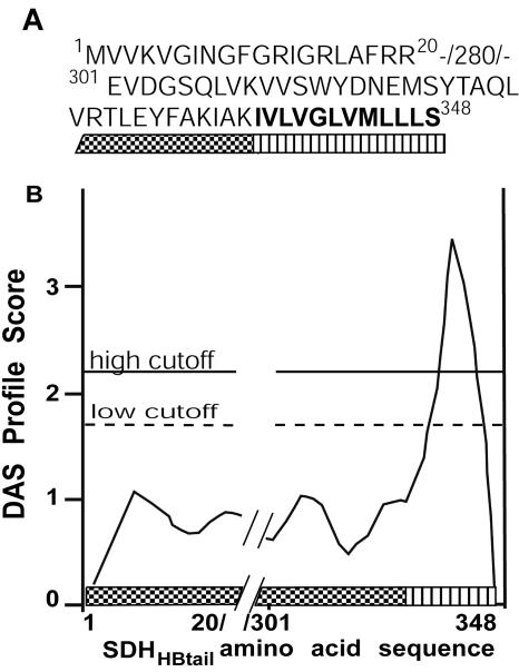
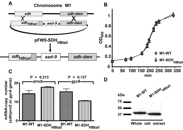
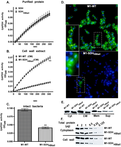
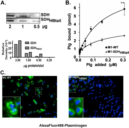
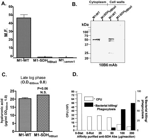
Surface dehydrogenase (SDH) is an anchorless, multifunctional protein displayed on the surfaces of group A Streptococcus (GAS) organisms. SDH is encoded by a single gene, sdh (gap or plr) that is essential for bacterial survival. Hence, the resulting nonfeasibility of creating a knockout mutant is a major limiting factor in studying its role in GAS pathogenesis. An insertion mutagenesis strategy was devised in which a nucleotide sequence encoding a hydrophobic tail of 12 amino acids ((337)IVLVGLVMLLLS(348)) was added at the 3' end of the sdh gene, successfully creating a viable mutant strain (M1-SDH(HBtail)). In this mutant strain, the SDH(HBtail) protein was not secreted in the medium but was retained in the cytoplasm and to some extent trapped within the cell wall. Hence, SDH(HBtail) was not displayed on the GAS surface. The mutant strain, M1-SDH(HBtail), grew at the same rate as the wild-type strain. The SDH(HBtail) protein displayed the same GAPDH activity as the wild-type SDH protein. Although the whole-cell extracts of the wild-type and mutant strains showed similar GAPDH activities, cell wall extracts of the mutant strain showed 5.5-fold less GAPDH activity than the wild-type strain. The mutant strain, M1-SDH(HBtail), bound significantly less human plasminogen, adhered poorly to human pharyngeal cells, and lost its innate antiphagocytic activity. These results indicate that the prevention of the cell surface export of SDH affects the virulence properties of GAS. The anchorless SDH protein, thus, is an important virulence factor.


Figures

FIG. 1.
FIG. 1.
Strategy to insert a hydrophobic tail at the C-terminal end of SDH. (A) Amino acid sequences of the SDH protein (only residues 1 to 20 and 301 to 336 are noted for brevity) and an inserted hydrophobic tail (337IVLVGLVMLLLS348; in bold) at the C-terminal end (sdh/plr in the M1 genome is annotated as SPy0274 [13]). The sequence of the hydrophobic tail for SDH was derived from that of the hydrophobic tail portion (2020IVLVGLGVMSLLLGMVLY2037) of the product of the epf gene (SPy0737) of the M1-SF370 genome (13). (B) DAS hydrophobicity index of the putative hydrophobic tail sequence (IVLVGLVMLLLS) to be inserted at the C-terminal end of the SDH molecule.
FIG. 2.
FIG. 2.
Growth characteristics of wild-type (M1-WT) and mutant (M1-SDHHBtail) GAS strains. (A) The mutant strain (M1-SDHHBtail) was derived from the wild-type strain (M1-SF370) using the pFW5-SDHHBtail suicide shuttle vector and by achieving double crossover during allelic-exchange events. (B) Comparison of the growth curves of the wild-type (M1-WT) (diamonds) and mutant (M1-SDHHBtail) (triangles) strains. (C) Comparison of the levels of sdh-specific mRNA expression with housekeeping gene (gyrA and proS)-specific mRNA expression in the late log phase of growth of the wild-type (M1-WT) and mutant (M1-SDHHBtail) strains by real-time quantitative PCR. P values were determined statistically using Student's t test. (D) Western immunoblotting of whole bacterial lysates (mutanolysin extract without raffinose buffer) of equal numbers of wild-type (M1-WT) and mutant (M1-SDHHBtail) strains using anti-SDH monoclonal antibody (13D5). Numbers on the left indicate positions of the molecular mass markers (kDa).
FIG. 3.
FIG. 3.
Surface and cell wall-associated GAPDH activities of the M1-WT and M1-SDHHBtail strains. The GAPDH activity of purified SDH, cell wall extract, or intact bacteria was measured by monitoring the conversion of G-3-P to 1,3-diphosphoglycerate spectrometrically (OD340) at different time intervals or as an endpoint reading after 5 min of incubation. (A) Comparison of GAPDH enzymatic activities of purified wild-type SDH (diamonds) and SDHHBtail (triangles) (3 μg of each). (B) Comparison of GAPDH enzyme activities of the cell wall extracts (CW) of the wild-type (M1-WT) (diamonds) and the mutant (M1-SDHHBtail) (triangles) strain (6 μg of each cell wall extract). (C) Comparison of GAPDH activities of intact M1-WT and M1-SDHHBtail GAS strains (200 μl of a bacterial suspension with an OD600 of 1.0, i.e.,108 CFU) as an endpoint reading of the supernatant obtained after 5 min of incubation in 600 μl of the reaction buffer followed by pelleting of the bacteria. Each reading point in panels A, B, and C represents an average from three independent experiments ± standard errors. (D) Indirect immunofluorescence microscopy of the wild-type (M1-WT) and mutant (M1-SDHHBtail) strains using affinity-purified monospecific rabbit anti-SDH IgG (Fab′ fragment) as the primary antibody followed by fluorescein isothiocyanate-labeled anti-rabbit Fab′-specific IgG (green fluorescence) as the secondary antibody. In parallel control experiments, both bacterial strains did not stain with the secondary conjugate antibody (not shown). The inset in each panel (dotted-line rectangle) is magnified (solid-line rectangle) to highlight bacterial-surface-associated SDH-specific punctate fluorescence. Note the absence of green fluorescence in the M1-SDHHBtail mutant strain. The blue fluorescence represents the DAPI-stained bacterial DNA. The background fluorescence in M1-SDHHBtail (inset) is due to lysed bacteria as revealed by the absence of DAPI stain. (E) Western blot analysis of the presence of SDH in culture supernatant (Sup) and subcellular fractions (CW, cell wall; Cyt, cytoplasm; and Mem, membrane) of the wild-type (M1-WT) and mutant (M1-SDHHBtail) strains using SDH-specific monoclonal antibody (13D5) (16). Both strains were grown to an OD of 0.8 in THY broth, and the supernatants were separated by centrifugation. The resulting bacterial pellets obtained from each strain were subjected to subcellular fractionation. Twenty-five percent-TCA-precipitated proteins obtained from 125 μl of the supernatant from each strain were tested for the presence of SDH. (F) Semiquantitative analysis of the presence of SDH in the cytoplasm and cell wall fractions of equal numbers of M1-WT and M1-SDHHBtail cells. Serial twofold dilutions of 4 μg of total proteins of the cytoplasmic and cell wall fractions obtained from both strains were resolved on PVDF membranes by SDS-PAGE and electroblotting. The presence of SDH was determined by immunoblotting using anti-SDH monoclonal antibody (13D5) and visualized with a chemiluminescence substrate.
FIG. 4.
FIG. 4.
Plasminogen-binding activities of the wild-type (M1-WT) and mutant (M1-SDHHBtail) GAS strains. (A) Plg-binding activities of serially diluted purified SDH and SDHHBtail (starting with 2 μg) proteins under native conditions using the slot blot device-based protein-ligand-binding method. Specific binding of Plg to purified SDH or SDHHBtail was determined with anti-human Plg antibody, visualized with a chemiluminescence substrate, and analyzed densitometrically. Bovine serum albumin was used as an internal control and showed no plasminogen binding (not shown). (B) Plg-binding activities of M1-WT and M1-SDHHBtail strains as measured by 96-well microtiter plate-based ligand-binding assays using intact bacteria (109 CFU) and various concentrations of AlexaFluor-488-labeled human Plg. The amount of bound Plg to bacterial strains was quantitated on the basis of a standard curve (fluorescence units versus the amount of labeled Plg). **, P < 0.001. (C) Fluorescence microscopy to reveal the direct Plg-binding ability of the wild-type (M1-WT) and the mutant (M1-SDHHBtail) strains using AlexaFluor-488-labeled human Plg (green fluorescence). The blue fluorescence represents the DAPI-stained bacterial DNA. The inset in each panel (dotted small rectangle) is magnified (solid-line rectangle) to highlight the Plg-binding pattern. Note the punctate Plg-binding pattern in the M1-WT strain versus the crescent-shaped and reduced Plg-binding pattern in the absence of the surface display of SDH in the M1-SDHHBtail strain.
FIG. 5.
FIG. 5.
Phagocytosis/bactericidal assays. (A) Growth of the wild-type (M1-WT) and mutant (M1-SDHHBtail) strains in human blood is expressed as a multiplication factor. The MF is the number of CFU obtained at the end of the incubation period over the number of CFU obtained at time zero. The M1Δemm1 strain was used as a negative control for antiphagocytic activity. Bars and error bars represent the means ± standard errors obtained from two independent experiments, each with triplicate samples. ***, P < 0.0001. (B) Western blot analysis of cell wall- and cytoplasm-associated proteins of the wild-type (M1-WT) and mutant (M1-SDHHBtail) strains using M1 protein-reacting 10B6 monoclonal antibody (mAb). Proteins were visualized with a chromogenic substrate. Each lane received a total of 5 μg of total cell wall-associated or cytoplasmic proteins. (C) Estimation of the hyaluronic capsule associated with the wild-type and mutant (M1-SDHHBtail) strains grown until late log phase (OD600 of 0.8). N.S., not significant (P > 0.05). (D) Opsonophagocytosis or inhibition of the antiphagocytic activity of the wild-type GAS strain in the presence of various concentrations of affinity-purified anti-SDH antibodies. The bactericidal assay was performed as described for panel A. The left y axis denotes the numbers of bacteria that survived at different concentrations of anti-SDH antibodies in the assay mixture at the end of the incubation period. The right y axis denotes percentages of bacteria being killed/phagocytosed at different concentrations of anti-SDH antibodies. Stat, stationary; Rot, rotation.
FIG. 6.
FIG. 6.
Bacterial adherence assays. Confluent human pharyngeal cells (Detroit 562) grown in 24-well tissue culture plates were incubated with the wild-type (M1-WT) and mutant (M1-SDHHBtail) strains (multiplicity of infection, 1:50, cells to bacteria) for 3 h. The mutant strain lacking the expression of the M1 protein (M1Δemm1) was used as an internal control in this assay. At the end of incubation, cell-associated bacteria were counted as numbers of CFU on sheep blood agar plates. The adherence index for each preparation was calculated as a percentage of the initial inoculum (4 × 10⁷ to 5 × 10⁷/well, i.e., 100 μl of an OD 1.0 bacterial suspension). Each error bar represents an average of results from three independent experiments, each from triplicate wells ± standard errors. **, P < 0.001. N.S., not significant.

Similar articles

Cited by

References

    1. Banks, D. J., S. F. Porcella, K. D. Barbian, S. B. Beres, L. E. Philips, J. M. Voyich, F. R. DeLeo, J. M. Martin, G. A. Somerville, and J. M. Musser. 2004. Progress toward characterization of the group A Streptococcus metagenome: complete genome sequence of a macrolide-resistant serotype M6 strain. J. Infect. Dis. 190:727-738. - PubMed
    1. Beres, S. B., G. L. Sylva, K. D. Barbian, B. Lei, J. S. Hoff, N. D. Mammarella, M.-Y. Liu, J. C. Smoot, S. F. Porcella, L. D. Parkins, D. S. Campbell, T. M. Smith, J. K. McCormick, D. Y. M. Leung, P. Schlievert, and J. M. Musser. 2002. Genome sequence of a serotype M3 strain of group A Streptococcus: phage-encoded toxins, the high-virulence phenotype, and clone emergence. Proc. Natl. Acad. Sci. USA 99:10078-10083. - PMC - PubMed
    1. Bergmann, S., M. Rohde, G. S. Chhatwal, and S. Hammerschmidt. 2001. Alpha-enolase of Streptococcus pneumoniae is a plasmin(ogen)-binding protein displayed on the bacterial cell surface. Mol. Microbiol. 40:1273-1287. - PubMed
    1. Biesecker, G., J. I. Harris, J. C. Thierry, J. E. Walker, and A. J. Wonacott. 1977. Sequence and structure of d-glyceraldehyde-3-phosphate dehydrogenase from Bacillus stearothermophilus. Nature 266:328-333. - PubMed
    1. Bisno, A. L., and D. L. Stevens. 1996. Streptococcal infections of skin and soft tissues. N. Engl. J. Med. 334:240-245. - PubMed

Publication types

MeSH terms

LinkOut - more resources