. 2005 Oct;73(10):6260-71.
doi: 10.1128/IAI.73.10.6260-6271.2005.

Identification of new secreted effectors in Salmonella enterica serovar Typhimurium

Affiliations

Identification of new secreted effectors in Salmonella enterica serovar Typhimurium

Kaoru Geddes et al. Infect Immun. 2005 Oct.

Abstract

A common theme in bacterial pathogenesis is the secretion of bacterial products that modify cellular functions to overcome host defenses. Gram-negative bacterial pathogens use type III secretion systems (TTSSs) to inject effector proteins into host cells. The genes encoding the structural components of the type III secretion apparatus are conserved among bacterial species and can be identified by sequence homology. In contrast, the sequences of secreted effector proteins are less conserved and are therefore difficult to identify. A strategy was developed to identify virulence factors secreted by Salmonella enterica serovar Typhimurium into the host cell cytoplasm. We constructed a transposon, which we refer to as mini-Tn5-cycler, to generate translational fusions between Salmonella chromosomal genes and a fragment of the calmodulin-dependent adenylate cyclase gene derived from Bordetella pertussis (cyaA'). In-frame fusions to bacterial proteins that are secreted into the eukaryotic cell cytoplasm were identified by high levels of cyclic AMP in infected cells. The assay was sufficiently sensitive that a single secreted fusion could be identified among several hundred that were not secreted. This approach identified three new effectors as well as seven that have been previously characterized. A deletion of one of the new effectors, steA (Salmonella translocated effector A), attenuated virulence. In addition, SteA localizes to the trans-Golgi network in both transfected and infected cells. This approach has identified new secreted effector proteins in Salmonella and will likely be useful for other organisms, even those in which genetic manipulation is more difficult.


Figures
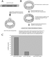
FIG. 1.
FIG. 1.
Schematic representation of the mini-Tn5-cycler transposon (A) and mutagenesis of srfH (B). IE and OE are the modified Tn5 transposon ends (also called mosaic ends [17]). cyaA′ is the promoterless 400 amino acids of the amino terminus of cyaA from Bordetella bronchiseptica (27, 40). KAN represents the Tn903 aminoglycoside phosphotransferase. To test our system, srfH was cloned into pBluescript (B). The purified plasmid was mutagenized in vitro using mini-Tn5-cycler transposon/transposase complexes (transposons) by the addition of 5 mM magnesium. Plasmids containing insertions were selected on LB-kanamycin plates, and in-frame insertions in srfH were identified by PCR and sequencing. An in-frame fusion of srfH-cyaA′ at the codon encoding amino acid 145 was then cloned into the suicide vector pKas32. The suicide vector containing an in-frame insertion of mini-Tn5-cycler was then electroporated into 14028s. Crossover events were selected on LB-kanamycin, and a double crossover was identified by PCR. (C) Secretion by srfH::mini-Tn5-cycler. Stationary-phase cultures of srfH::mini-Tn5-cycler, a mutant expressing a fusion from a low-copy-number plasmid (psrfH-cyaA′), or a mutant expressing a fusion of CyaA′ to the β-galactosidase alpha peptide (placZ-cyaA′) were used to infect J774 macrophages for 8 h at an MOI of ∼1. Infected cells were lysed with 0.1 M HCl, and the concentration of cAMP (pmol/ml) in the lysates was measured by ELISA.
FIG. 2.
FIG. 2.
Strategy for identifying effectors. Libraries of approximately 5,000 insertions were generated by electroporating mini-Tn5-cycler transposon/transposase complexes into either 14028s or a ΔslrP derivative. These colonies were diluted in LB broth to approximately 500 to 1,000 bacteria/ml (based on optical density readings at 600 nm), and 100-μl aliquots of the diluted library were grown to either stationary phase or late log phase. These cultures were then used to infect J774 cells or HeLa cells seeded in 96-well plates. After 8 to 10 h of infection for J774 cells or 2 h for HeLa cells, the medium was removed, and 0.1 M HCl was added to lyse the cells. The level of cAMP in each well was then measured using an ELISA. Any pool from which infected cells showed at least 10 times higher levels of cAMP than the background level was isolated and screened further by repooling into smaller groups (10 or 1) until individual bacteria containing the cognate CyaA′ fusion were obtained.
FIG. 3.
FIG. 3.
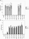
SPI-1 TTSS (A)- and SPI-2 TTSS (B)-dependent secretion of cyaA′ fusions into J774 cells. Levels of cAMP were measured within the macrophage-like cell line J774 following infection with the 10 cyaA′ fusions listed in Table 2. Three different backgrounds were used for this experiment. These were WT (gray bars), invA::cat (black bars), and ssaK::cat (white bars). Bacteria were grown to late log phase and used to infect J774 cells at an input MOI of ∼50 for 1 hour (A) or were grown to stationary phase and used to infect J774 cells for 8 h at an input MOI of ∼250 (B). The cells were then lysed with 0.1 M HCl, and an ELISA (Assay Designs) was used to quantitate the cAMP levels. The cAMP concentration (in pmol/ml) was measured in triplicate samples, and the error bars represent 1 standard error of the mean.
FIG. 4.
FIG. 4.
SPI-1 TTSS- and SPI-2 TTSS-dependent secretion of full-length CyaA′ fusions. Levels of cAMP were measured within the macrophage-like cell line J774 following infection with SteA-CyaA′, SteB-CyaA′, and SteC-CyaA′. Three different backgrounds were used for this experiment. These were WT (gray bars), invA::cat (black bars), and ssaK::cat (white bars). Bacteria were grown to late log phase and used to infect J774 cells at an input MOI of ∼50 for 1 hour (SPI-1 conditions) or were grown to stationary phase and used to infect J774 cells for 8 h at an input MOI of ∼250 (SPI-2 conditions). The cells were then lysed with 0.1 M HCl, and an ELISA (Assay Designs) was used to quantitate the cAMP levels. The cAMP concentration (in pmol/ml) was measured in triplicate samples, and the error bars represent 1 standard error of the mean.
FIG. 5.
FIG. 5.
A SteA-EGFP fusion expressed in HeLa cells colocalizes with the TGN. HeLa cells were transfected for 24 h with pEGFP (bottom panels) or pSteA-EGFP (top panels), and Bodipy-TR-ceramide (red) was used to stain the TGN. The images shown are 0.2-μm z sections captured using deconvolution microscopy. EGFP fluorescence images are shown in panels A and D, and Bodipy-TR-ceramide fluorescence images are shown in panels B and E. Panels C and F show mergers of panels A and B and panels D and E, respectively. In panels C and F, the areas where EGFP and Bodipy-TR-ceramide fluorescence overlap are yellow.
FIG. 6.
FIG. 6.
Secreted SteA colocalizes with the TGN in infected HeLa cells. HeLa cells were infected with SteA-HA/14028s (top panels) or with WT 14028s (bottom panels) for 4 hours at an MOI of 100 under SPI-1-inducing conditions. HA-specific antibodies were used to visualize HA-tagged SteA (green). Bodipy-TR-ceramide (red) was used to stain the TGN, and the DNA stain DRAQ5 (blue) was used to visualize host cell nuclei and bacteria. DRAQ5 fluorescence images are shown in panels A and E, HA tag-specific fluorescence images are shown in panels B and F, and Bodipy-TR-ceramide fluorescence images are shown in panels C and G. Panels D and H show mergers of the images from panels A to C and E to G, respectively.

References

    1. Ausubel, F. M. 1987. Current protocols in molecular biology. Greene Publishing Associates, Brooklyn, N.Y.
    1. Bajaj, V., R. L. Lucas, C. Hwang, and C. A. Lee. 1996. Co-ordinate regulation of Salmonella typhimurium invasion genes by environmental and regulatory factors is mediated by control of hilA expression. Mol. Microbiol. 22:703-714. - PubMed
    1. Brazilian National Genome Project Consortium. 2003. The complete genome sequence of Chromobacterium violaceum reveals remarkable and exploitable bacterial adaptability. Proc. Natl. Acad. Sci. USA 100:11660-11665. - PMC - PubMed
    1. Chakravortty, D., I. Hansen-Wester, and M. Hensel. 2002. Salmonella pathogenicity island 2 mediates protection of intracellular Salmonella from reactive nitrogen intermediates. J. Exp. Med. 195:1155-1166. - PMC - PubMed
    1. Chen, J., K. S. de Felipe, M. Clarke, H. Lu, O. R. Anderson, G. Segal, and H. A. Shuman. 2004. Legionella effectors that promote nonlytic release from protozoa. Science 303:1358-1361. - PubMed

Publication types

MeSH terms

LinkOut - more resources