. 2005 Oct;73(10):6299-310.
doi: 10.1128/IAI.73.10.6299-6310.2005.

Infection with Mycobacterium ulcerans induces persistent inflammatory responses in mice

Affiliations

Infection with Mycobacterium ulcerans induces persistent inflammatory responses in mice

Martinha S Oliveira et al. Infect Immun. 2005 Oct.

Abstract

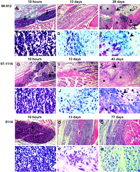
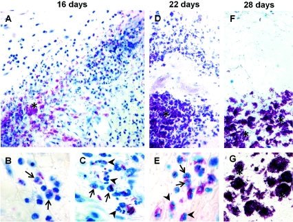
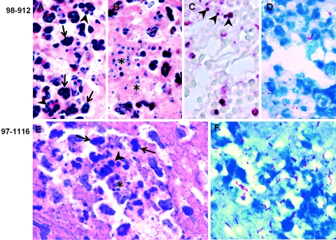
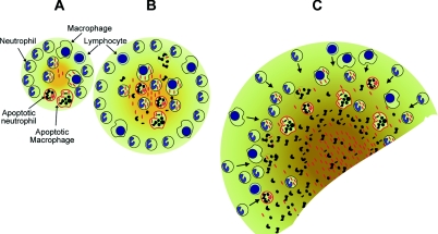
Buruli ulcer (BU) is a devastating, necrotizing, tropical skin disease caused by infections with Mycobacterium ulcerans. In contrast to other mycobacterioses, BU has been associated with minimal or absent inflammation. However, here we show that in the mouse M. ulcerans induces persistent inflammatory responses with virulence-dependent patterns. Mycolactone-positive, cytotoxic strains are virulent for mice and multiply progressively, inducing both early and persistent acute inflammatory responses. The cytotoxicity of these strains leads to progressive destruction of the inflammatory infiltrates by postapoptotic secondary necrosis, generating necrotic acellular areas with extracellular bacilli released by the lysis of infected phagocytes. The necrotic areas, always surrounded by acute inflammatory infiltrates, expand through the progressive invasion of healthy tissues around the initial necrotic lesions by bacteria and by newly recruited acute inflammatory cells. Our observations show that the lack of inflammatory infiltrates in the extensive areas of necrosis seen in advanced infections results from the destruction of continuously produced inflammatory infiltrates and not from M. ulcerans-induced local or systemic immunosuppression. Whether this is the mechanism behind the predominance of minimal or absent inflammatory responses in BU biopsies remains to be elucidated.


Figures

FIG. 1.
FIG. 1.
Cytotoxicity of M. ulcerans clinical isolates to BMDM. BMDM were not infected (A) or were infected with M. ulcerans strain 5114 (B), 97-1116 (C), or 98-912 (D and E) at an MOI of 1:1. Macrophages were photographed 4 days after infection by phase-contrast microscopy (A to D) or with fluorescence microscopy for samples stained with TUNEL (E). Positive TUNEL staining (yellow-green fluorescent nuclei in E) points to the apoptotic nature of cell death induced by the cytotoxic strain 98- 912. Bars, 50 μm.
FIG. 2.
FIG. 2.
Footpad swelling and proliferation of different strains of M. ulcerans. Panels A and C show swelling and AFB counts, respectively, of BALB/c mice footpads infected subcutaneously with 5.0 log10 AFB of M. ulcerans 98-912 (▪), 5.3 log10 AFB of M. ulcerans 97-1116 (▴), or 5.1 log10 AFB of M. ulcerans 5114 (•). Panel B shows swelling of footpads infected with 5.5 log10 AFB (▪) or 2.5 log10 AFB (□) of M. ulcerans 98-912 or 5.5 log10 AFB of M. ulcerans 5114 (•). Footpad swelling was measured in eight mice from each group. Significant differences in footpad swelling were calculated by comparing infected and noninfected mice. Significant differences in AFB counts were determined by comparing the bacterial loads of five footpads from each group with the respective initial inoculum. Calculations were performed using Student's t test (*, P < 0.05; **, P < 0.01; ***, P < 0.001). Mice were sacrificed for ethical reasons after the emergence of ulceration (†). Results are from one representative of three independent experiments. Error bars indicate standard deviations.
FIG. 3.
FIG. 3.
Kinetics of recruitment of macrophages and neutrophils to the peritoneal cavity following infection with M. ulcerans. BALB/c mice were infected intraperitoneally with 6.1 log10 AFB of M. ulcerans 98-912 (▪) or 6.2 log10 AFB of M. ulcerans 5114 (•). Peritoneal cells were recovered by peritoneal lavage, and the total number of leukocytes was determined. Counting of neutrophils (solid lines) and macrophages (dashed lines) was performed on Hemacolor-stained cytospins. *, P < 0.05; ***, P < 0.001 (determined by Student's t test, comparing infected mice with mice inoculated with PBS). Results are from one representative experiment of two independent experiments. Error bars indicate standard deviations.
FIG. 4.
FIG. 4.
Histological sections of footpads of mice infected with M. ulcerans. BALB/c mice were infected with 5.9 log10 AFB of M. ulcerans 98-912 (A to F), 5.5 log10 AFB of M. ulcerans 97-1116 (G to L), or 5.8 log10 AFB of M. ulcerans 5114 (M to R). Footpads were collected at the indicated times postinfection, and tissue sections were stained with HE (A, B, C, E, G, H, I, K, M, N, O, and Q) or ZN (D, F, J, L, P, and R). Magnifications, ×60 (A, C, E, G, I, K, M, O, and Q) and ×350 (B, D, F, H, J, L, N, P, and R). Panels A, B, G, H, M, and N show an acute inflammatory response with predominance of neutrophils (arrows) early after infection by all strains of M. ulcerans. Panels C to F and I to L depict advanced stages of infection with M. ulcerans 98-912 or 97-1116, respectively, and show necrosis of the central focus of infection (E, F, K, and L, asterisks) and high numbers of extracellular bacteria (F and L), surrounded by a band of acute cellular infiltrate (E and K, arrowheads). In contrast, advanced stages of infection with M. ulcerans 5114 show predominantly mononuclear infiltrates with intracellular bacilli, epithelioid transformation (O to R), and granuloma-like organization (P). Results are from one representative experiment of two independent experiments.
FIG. 5.
FIG. 5.
Histological sections of lesions at late stages of infection with a mouse-virulent M. ulcerans strain. Footpads of mice infected with 5.9 log10 AFB of M. ulcerans 98-912 were analyzed at 16 (A, B, and C), 22 (D and E), or 28 (F and G) days postinfection. Tissue sections were stained with ZN. Magnifications, ×250 (A, D, and F) and ×680 (B, C, E, and G). Panels A, D, and F show acellular necrotic areas that are extensive in advanced infections (F), associated with extracellular bacilli (asterisks), surrounded by a band of acute inflammatory cellular infiltrate (A and D); the acute infiltrate at the periphery of the necrotic area in F is outside the micrograph. Higher magnifications of the acute infiltrates in A and D show intracellular bacilli (arrows in panels B, C, and E) and leukocyte death with pyknosis and karyorrhexis (arrowheads in panels C and E).
FIG. 6.
FIG. 6.
Histological sections of inflammatory infiltrates at early stages of infection with mouse-virulent M. ulcerans strains. Mouse footpads were inoculated with 5.9 log10 AFB of M. ulcerans 98-912 (A to D) or 5.5 log10 AFB of M. ulcerans 97-1116 (E and F) for 24 h (A to D) or 48 h (E and F). Serial tissue sections (A to D and E and F) were stained with HE (A, B, and E), TUNEL (C), or ZN (D and F). Magnifications, ×750 (C), ×1,000 (A, B, and E), and ×1,250 (D and F). These panels show leukocyte cell death colocalized with bacilli in the acute inflammatory infiltrate (D and F), with evidence of pyknosis (A and E, arrows), karyorrhexis (A, C, and E, arrowheads), and cellular debris (B and E, asterisks). TUNEL staining (red nuclei and nuclear fragments in C) points to the apoptotic nature of cell death.
FIG. 7.
FIG. 7.
Schematic representation of the progression of BU. Panel A shows M. ulcerans (red bacilli) being phagocytosed in the initial stage of infection by neutrophils and macrophages that are present in the early acute inflammatory infiltrate. Panel B represents a more advanced infection stage characterized by the onset of cell destruction, showing predominant neutrophilic cell infiltrate, apoptotic neutrophils and macrophages, and cell debris resulting from postapoptotic secondary necrosis. Panel C highlights an advanced stage of BU with extensive acellular areas with numerous extracellular bacilli, tissue damage represented by accumulation of cellular fragments, continued predominance of neutrophils, and low number of macrophages and lymphocytes. This stage is reminiscent of what has been observed in biopsies of advanced BU. The orange-yellow gradient represents the production and spreading of M. ulcerans exotoxin.

References

    1. Abalos, F. M., J. Aguiar, Sr., A. Guedenon, F. Portaels, and W. M. Meyers. 2000. Mycobacterium ulcerans infection (Buruli ulcer): a case report of the disseminated nonulcerative form. Ann. Diagn. Pathol. 4:386-390. - PubMed
    1. Appelberg, R., J. M. Pedrosa, and M. T. Silva. 1991. Host and bacterial factors control the Mycobacterium avium-induced chronic peritoneal granulocytosis in mice. Clin. Exp. Immunol. 83:231-236. - PMC - PubMed
    1. Busam, K. J., T. E. Kiehn, S. P. Salob, and P. L. Myskowski. 1999. Histologic reactions to cutaneous infections by Mycobacterium haemophilum. Am. J. Surg. Pathol. 23:1379-1385. - PubMed
    1. Chemlal, K., K. De Ridder, P. A. Fonteyne, W. M. Meyers, J. Swings, and F. Portaels. 2001. The use of IS2404 restriction fragment length polymorphisms suggests the diversity of Mycobacterium ulcerans from different geographical areas. Am. J. Trop. Med. Hyg. 64:270-273. - PubMed
    1. Chemlal, K., G. Huys, P. A. Fonteyne, V. Vincent, A. G. Lopez, L. Rigouts, J. Swings, W. M. Meyers, and F. Portaels. 2001. Evaluation of PCR-restriction profile analysis and IS2404 restriction fragment length polymorphism and amplified fragment length polymorphism fingerprinting for identification and typing of Mycobacterium ulcerans and M. marinum. J. Clin. Microbiol. 39:3272-3278. - PMC - PubMed

Publication types