. 2005 Oct;73(10):6372-82.
doi: 10.1128/IAI.73.10.6372-6382.2005.

Live nonpathogenic parasitic vector as a candidate vaccine against visceral leishmaniasis

Affiliations

Live nonpathogenic parasitic vector as a candidate vaccine against visceral leishmaniasis

Marie Breton et al. Infect Immun. 2005 Oct.

Abstract

To date, there are no proven vaccines against any form of leishmaniasis. The development of live attenuated vectors shows promise in the field of Leishmania vaccination because these organisms mimic more effectively the course of real infections and can elicit potent activation of the immune system. In the present study, we investigated the potential of a parasitic protozoan that is nonpathogenic to humans, Leishmania tarentolae, as a live candidate vaccine that efficiently targets dendritic cells and lymphoid organs, thus enhancing antigen presentation and consequently influencing the magnitude and quality of T-cell immune responses. We demonstrated that L. tarentolae activates the dendritic cell maturation process and induces T-cell proliferation and the production of gamma interferon, thus skewing CD4(+) T cells toward a Th1 cell phenotype. More importantly, we found that a single intraperitoneal injection of L. tarentolae could elicit a protective immune response against infectious challenge with Leishmania donovani in susceptible BALB/c mice. These results suggest that the use of L. tarentolae as a live vaccine vector may represent a promising approach for improving the effectiveness and safety of candidate live vaccines against Leishmania infections and possibly other intracellular pathogens for which T-cell mediated responses are critical for the development of protective immunity.


Figures

FIG. 1.
FIG. 1.
L. tarentolae uptake by murine and human macrophages. Murine monocytic cells, murine monocyte-derived macrophages, and the human monocytic cell line TPH1 were used to evaluate the ability of L. tarentolae to infect cells in vitro. The ability of L. tarentolae to enter intraperitoneal macrophages was evaluated in vivo. (A) Giemsa staining of J774 infected macrophages with L. tarentolae. (B) J774 macrophages stained with Evans blue and viewed by confocal microscopy. (C) FACS evaluation of the percentage of GFP-expressing L. tarentolae-infected macrophages 24 h following infection. (D) Giemsa staining of L. tarentolae-infected intraperitoneal macrophages. (E) Giemsa staining of THP1 macrophages infected with L. tarentolae 24 h following infection. (F) FACS evaluation of the percentage of GFP-expressing L. tarentolae-infected THP1 macrophages 24 h postinfection. (G) Percentage of THP1 macrophages infected by L. tarentolae as monitored by microscopic evaluation of Giemsa-stained tissues at different times following infection. On average, 200 macrophages per time were counted. (H) FACS evaluation of the percentage of monocyte-derived macrophages infected with GFP-expressing L. tarentolae, as determined by cytokine differentiation of CD14 purification of human PBMCs 24 h postinfection.
FIG. 2.
FIG. 2.
L. tarentolae uptake by iMDDCs. Target cells were obtained by cytokine differentiation (IL-4, granulocyte-macrophage colony-stimulating factor) of CD14 purified from human PBMCs by MACS magnetic bead isolation. The ability of L. tarentolae to enter these cells was evaluated by adding GFP-expressing L. tarentolae to iMDDCs at a parasite/macrophage ratio of 10:1. The infection was monitored 12 h after infection by FACS. (A) Confocal microscopy of infected iMDDCs. (B) Flow cytometry profile showing the percentage of infected iMDDCs. (C) Expression of activation markers 48 h following in vitro stimulation of iMDDCs with L. tarentolae (Ltar) at a macrophage/parasite ratio of 10:1 or with 100 ng of LPS. The expression of the different surface markers was evaluated by FACS analysis by incubating the cells with the following monoclonal antibodies: anti-HLADR, anti-CD40, anti-CD80 (B7.1), anti-CD86 (B7.2), and anti-CD83.
FIG. 3.
FIG. 3.
Leukocyte accumulation in the air pouch mouse system in response to Leishmania inoculation. Air pouches were raised on the dorsum of 6- to 8-week-old female CD-1 mice. One milliliter of endotoxin-free PBS with or without LPS or Leishmania (5 × 10⁷ cells) was injected into the pouches, and the exudates were collected 6 h after inoculation. The nonpathogenic species L. tarentolae (Ltar) and the pathogenic species L. major (Lm) were used in these studies. (A) Number of leukocytes, as determined microscopically with a hemacytometer. (B) Sum of the recruited neutrophils, monocytes/macrophages, eosinophils, and lymphocytes, as determined on slides by microscopic analysis using Cytospin stained with a Diff-Quick solution. The proportion of each cell type was determined by examining 300 cells. The data represent the means ± standard errors of two independent experiments (five mice each). Asterisks and daggers indicate statistically significant differences between experimental mice and the PBS control mice (asterisks, P < 0.01; daggers, P < 0.05).
FIG. 4.
FIG. 4.
L. tarentolae induces T-cell proliferation and IFN-γ production in mice. (A) Proliferation of lymphocytes from BALB/c mice previously infected with L. tarentolae. A total of 5 × 10⁵ splenocytes were isolated from immunized BALB/c mice at 1, 2, 4, 8, and 12 weeks postinfection, grown in culture, and restimulated for 4 days with L. tarentolae live promastigotes (Ltar). Three days following stimulation, 1 μCi of [3H]thymidine was added to each culture for 24 h, and then [3H]thymidine incorporation was measured as described in Materials and Methods. (B) IFN-γ cytokine production by the splenocytes at different times following vaccination of mice with 5 × 10⁶ L. tarentolae cells. Levels of IFN-γ were measured by a sandwich ELISA in culture supernatants of splenocytes 4 days following in vitro restimulation with L. tarentolae promastigotes. The data are the means ± standard errors for levels of cytokine production in three independent experiments (five mice each). Asterisks indicate that the P value is <0.01 for a comparison of stimulated and unstimulated splenocytes.
FIG. 5.
FIG. 5.
Protective immunity in mice preimmunized with L. tarentolae following a challenge with a virulent L. donovani strain. Six weeks after immunization with L. tarentolae, 5 × 10⁷ stationary-phase LUC-expressing L. donovani promastigotes (Ld) were injected into the tail vein of BALB/c mice. Naïve control mice were treated similarly. At 1 month postchallenge, the spleen and the liver were harvested, and the luciferase activity was measured as an indicator of the presence of parasites. The data are the means ± standard errors for five mice per group and are representative of three experiments in which similar results were obtained. Asterisks and daggers indicate statistically significant differences between experimental mice and control mice (asterisks, P < 0.01; daggers, P < 0.05).
FIG. 6.
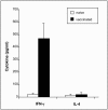
FIG. 6.
Cytokines produced by BALB/c mice initially infected with L. tarentolae after challenge with L. donovani. The IFN-γ and IL-4 cytokine levels produced by splenocytes nearly 12 weeks following vaccination with 5 × 10⁶ L. tarentolae and 6 weeks following challenge with 5 × 10⁷ L. donovani were determined. Cytokines were measured by an ELISA by using culture supernatants, as described in the legend to Fig. 4, except that the experiments were conducted 6 weeks after challenge with L. donovani. The bars indicate the mean cytokine production of three independent experiments (five mice each), and the error bars indicate standard errors.

References

    1. Abdelhak, S., H. Louzir, J. Timm, L. Blel, Z. Benlasfar, M. Lagranderie, M. Gheorghiu, K. Dellagi, and B. Gicquel. 1995. Recombinant BCG expressing the leishmania surface antigen Gp63 induces protective immunity against Leishmania major infection in BALB/c mice. Microbiology 141:1585-1592. - PubMed
    1. Ahmed, S., M. Colmenares, L. Soong, K. Goldsmith-Pestana, L. Munstermann, R. Molina, and D. McMahon-Pratt. 2003. Intradermal infection model for pathogenesis and vaccine studies of murine visceral leishmaniasis. Infect. Immun. 71:401-410. - PMC - PubMed
    1. Alexander, J., G. H. Coombs, and J. C. Mottram. 1998. Leishmania mexicana cysteine proteinase-deficient mutants have attenuated virulence for mice and potentiate a Th1 response. J. Immunol. 161:6794-6801. - PubMed
    1. Ashford, R. W. 2001. Current usage of nomenclature for parasitic diseases, with special reference to those involving arthropods. Med. Vet. Entomol. 15:121-125. - PubMed
    1. Athman, R., and D. Philpott. 2004. Innate immunity via Toll-like receptors and Nod proteins. Curr. Opin. Microbiol. 7:25-32. - PubMed

Publication types