. 2005 Oct;73(10):6437-45.
doi: 10.1128/IAI.73.10.6437-6445.2005.

Acid-induced activation of the urease promoters is mediated directly by the ArsRS two-component system of Helicobacter pylori

Affiliations

Acid-induced activation of the urease promoters is mediated directly by the ArsRS two-component system of Helicobacter pylori

Michael Pflock et al. Infect Immun. 2005 Oct.

Abstract

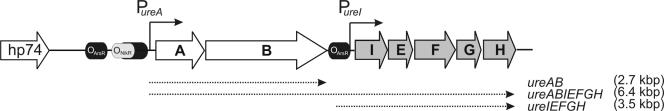
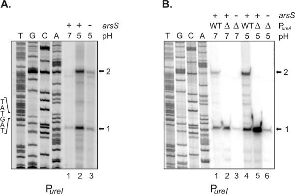
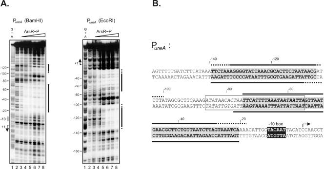
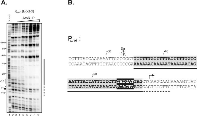
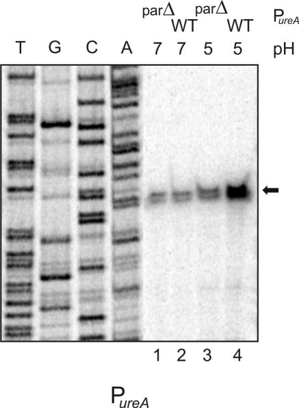
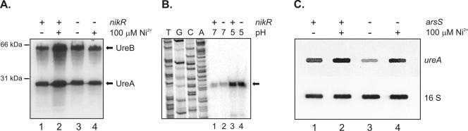
The nickel-containing enzyme urease is an essential colonization factor of the human gastric pathogen Helicobacter pylori which enables the bacteria to survive the low-pH conditions of the stomach. Transcription of the urease genes is positively controlled in response to increasing concentrations of nickel ions and acidic pH. Here we demonstrate that acid-induced transcription of the urease genes is mediated directly by the ArsRS two-component system. Footprint analyses identify binding sites of the phosphorylated ArsR response regulator within the ureA and ureI promoters. Furthermore, deletion of a distal upstream ArsR binding site of the ureA promoter demonstrates its role in acid-dependent activation of the promoter. In addition, acid-induced transcription of the ureA gene is unaltered in a nikR mutant, providing evidence that pH-responsive regulation and nickel-responsive regulation of the ureA promoter are mediated by independent mechanisms involving the ArsR response regulator and the NikR protein.


Figures

FIG. 1.
FIG. 1.
Schematic representation of the urease operon. The urease genes and the upstream ORF HP0074 (hp74) encoding a lipoprotein signal peptidase (32) are shown, and the directions of transcription of these genes are indicated by arrows. The PureA and PureI promoters are marked by thin arrows. The gray oval upstream of PureA indicates the operator of the NikR regulator mapped by Delany et al. (13). The black ovals indicate the binding sites of the response regulator ArsR. Dotted arrows represent the transcripts whose synthesis is directed by the PureA and PureI promoters, respectively, and which are further processed by endonucleolytic cleavage (1). The sizes of these transcripts are indicated on the right. The figure is not drawn to scale.
FIG. 2.
FIG. 2.
Analysis of transcription from the PureI promoter in H. pylori strains grown at neutral pH and exposed to pH 5.0. A. Primer extension experiments using the radiolabeled oligonucleotide ureIPE were performed on equal amounts of RNAs extracted from H. pylori G27 grown at neutral pH (lane 1) and from strains G27 and G27/HP165::km which were exposed to pH 5.0 for 60 min (lanes 2 and 3, respectively). The elongated primer products 1 and 2 are indicated by arrows. The sequence of the −10 element of the PureI promoter is given on the left. The sequencing ladders (lanes T, G, C, and A) were obtained by annealing primer ureIPE to plasmid pSL-ureI. B. Primer extension experiments with oligonucleotide ureIPE were performed on equal amounts of RNAs extracted from H. pylori G27 (lanes 1 and 4), G27/ΔPureA (lanes 2 and 5), and G27/ΔarsSΔPureA (lanes 3 and 6) grown at neutral pH or exposed to pH 5.0 for 60 min. WT, wild type.
FIG. 3.
FIG. 3.
Binding of ArsR∼P to the PureA promoter. A. DNase I footprint experiments were performed on a 339-bp EcoRI-BamHI fragment containing the PureA promoter which was end labeled at the BamHI and EcoRI termini, respectively, by adding increasing amounts of His6-ArsR phosphorylated in vitro by acetylphosphate. In lanes 2 to 8, His6-ArsR is present at concentrations of 0, 0.75, 1.5, 3.0, 4.5, 6.0, and 7.5 μM, respectively. The numbers on the left indicate nucleotide positions with respect to the transcriptional start site, which is marked by an arrow. The open bar indicates the position of the −10 promoter element. The solid and broken bars on the right indicate the minimum and maximum regions of DNase I protection, respectively. Lane 1 contained a G+A sequence reaction mixture with the DNA probe used as a size marker (22). B. Schematic representation of the PureA promoter. The −10 promoter element is highlighted by black shading, and the transcriptional start site is indicated by an arrow above the double-stranded sequence. Black bars below and above the sequence indicate the minimum (solid lines) and maximum (dashed lines) regions protected from DNase I digestion by the binding of ArsR∼P to the PureA promoter probe labeled at the BamHI and EcoRI termini, respectively. The overlapping regions which were clearly protected on both probes are highlighted by gray shading. The NikR binding site mapped by Delany et al. (13) is boxed, and the respective sequence motif is in italics. Numbers above the sequence indicate the nucleotide position with respect to the transcriptional start site (+1).
FIG. 4.
FIG. 4.
Binding of ArsR∼P to the PureI promoter. A. A DNase I footprint experiment was performed on a 414-bp BamHI-EcoRI fragment containing the PureI promoter which was end labeled at the EcoRI terminus. In lanes 2 to 9, His6-ArsR which was phosphorylated in vitro by acetylphosphate is present at concentrations of 0, 0.37, 0.75, 1.5, 3.0, 4.5, 6.0, and 7.5 μM, respectively. The numbers on the left indicate nucleotide positions with respect to the transcriptional start site, which is marked by an arrow. The open bar indicates the position of the −10 promoter element. The solid and broken bars on the right indicate the minimum and maximum regions of DNase I protection, respectively. Lane 1 contained a G+A sequence reaction mixture with the DNA probe used as a size marker (22). B. Schematic representation of the PureI promoter. The −10 promoter element is highlighted by black shading, and the transcriptional start site is indicated by an arrow above the double-stranded sequence. The black bars below the sequence indicate the minimum (solid lines) and maximum (dashed lines) regions protected from DNase I digestion by the binding of ArsR∼P to the PureI promoter probe. The minimum region of protection is also highlighted by gray shading. Numbers above the sequence indicate the nucleotide position with respect to the transcriptional start site (+1). The scissors above the sequence indicate the 5′ end of the ureI-specific transcript observed in the primer extension experiments whose synthesis is directed by the PureA promoter.
FIG. 5.
FIG. 5.
Analysis of ureA expression in H. pylori strains G27 and G27/parΔPureA by primer extension analysis. Primer extension experiments using the radiolabeled oligonucleotide ureAPE were performed on equal amounts of RNAs extracted from H. pylori G27 grown at neutral pH (lane 2) and pH 5.0 (lane 4) and from strain G27/parΔPureA grown at neutral pH (lane 1) and at pH 5.0 (lane 3). The arrow on the right indicates the position of the cDNA corresponding to the ureA-specific transcript. The sequencing ladders (lanes T, G, C, and A) were obtained by annealing primer ureAPE to plasmid pSLparΔPureA.
FIG. 6.
FIG. 6.
Analysis of the expression of urease in H. pylori strains G27, G27/nikR::km and G27/HP165::km. A. Immunoblot analysis of equal amounts of whole-cell protein prepared from H. pylori G27 grown in brucella broth (lane 1) or brucella broth supplemented with 100 μM NiCl2 (lane 2) and from G27/nikR::km grown in brucella broth (lane 3) or brucella broth supplemented with 100 μM NiCl2 (lane 4) with a polyclonal antiserum directed against H. pylori urease. The UreB and UreA proteins are indicated on the right, and the positions of molecular mass markers are given on the left. B. Primer extension analysis using the radiolabeled oligonucleotide ureAPE was performed on equal amounts of RNAs extracted from H. pylori G27 grown at neutral pH (lane 1) and exposed to pH 5.0 (lane 3) and from G27/nikR::km grown at neutral pH (lane 2) and exposed to pH 5.0 (lane 4). The arrow on the right indicates the position of the cDNA corresponding to the ureA-specific transcript. C. Slot blot Northern hybridization performed with RNAs extracted from H. pylori G27 grown in brucella broth (lane 1) or brucella broth supplemented with 100 μM NiCl2 (lane 2) and from G27/HP165::km grown in brucella broth (lane 3) or brucella broth supplemented with 100 μM NiCl2 (lane 4). Hybridization was performed with ureA- and 16S rRNA-specific probes as indicated on the right.

References

    1. Akada, J. K., M. Shirai, H. Takeuchi, M. Tsuda, and T. Nakazawa. 2000. Identification of the urease operon in Helicobacter pylori and its control by mRNA decay in response to pH. Mol. Microbiol. 36:1071-1084. - PubMed
    1. Bauerfeind, P., R. Garner, B. E. Dunn, and H. L. Mobley. 1997. Synthesis and activity of Helicobacter pylori urease and catalase at low pH. Gut 40:25-30. - PMC - PubMed
    1. Beier, D., G. Spohn, R. Rappuoli, and V. Scarlato. 1997. Identification and characterization of an operon of Helicobacter pylori that is involved in motility and stress adaptation. J. Bacteriol. 179:4676-4683. - PMC - PubMed
    1. Beier, D., and R. Frank. 2000. Molecular characterization of two-component systems of Helicobacter pylori. J. Bacteriol. 182:2068-2076. - PMC - PubMed
    1. Bijlsma, J. J. E., M. M. Gerrits, R. Imamdi, C. M. J. E. Vandenbroucke-Grauls, and J. G. Kusters. 1998. Urease-positive, acid-sensitive mutants of Helicobacter pylori: urease-independent acid resistance involved in growth at low pH. FEMS Microbiol. Lett. 167:309-313. - PubMed

Publication types

MeSH terms

LinkOut - more resources