. 2005 Oct;73(10):6674-9.
doi: 10.1128/IAI.73.10.6674-6679.2005.

Hyperinfectivity of human-passaged Vibrio cholerae can be modeled by growth in the infant mouse

Affiliations

Hyperinfectivity of human-passaged Vibrio cholerae can be modeled by growth in the infant mouse

Ashfaqul Alam et al. Infect Immun. 2005 Oct.

Abstract

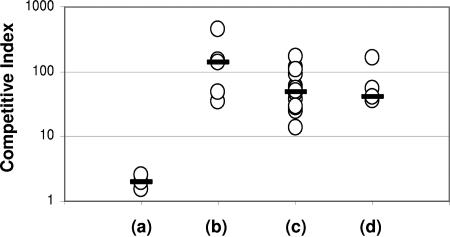
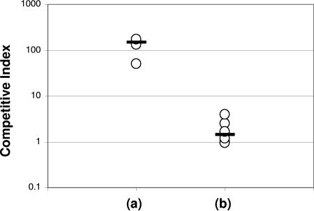
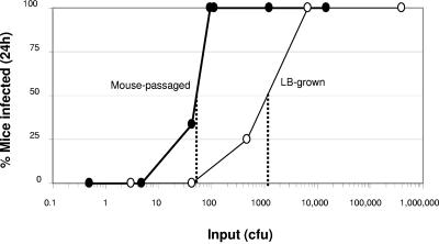
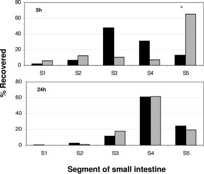
It has previously been shown that passage of Vibrio cholerae through the human intestine imparts a transient hyperinfectious phenotype that may contribute to the epidemic spread of cholera. The mechanism underlying this human-passaged hyperinfectivity is incompletely understood, in part due to inherent difficulties in recovering and studying organisms that are freshly passed in human stool. Here, we demonstrate that passage of V. cholerae through the infant mouse intestine leads to an equivalent degree of hyperinfectivity as passage through the human host. We have used this infant mouse model of host-passaged hyperinfectivity to characterize the timing and the anatomic location of the competitive advantage of mouse-passaged V. cholerae as well as the contribution of three type IV pili to the phenotype.


Figures

FIG. 1.
FIG. 1.
Competition assays in infant mice between LacZ+ V. cholerae N16961 and LacZ V. cholerae KFV10 grown in various conditions prior to competition in CD-1 infant mice. (a) LacZ+ versus LacZ V. cholerae, both grown to stationary phase in LB; (b) mouse-passaged LacZ+ V. cholerae versus LacZ V. cholerae grown to stationary phase in LB; (c) mouse-passaged LacZ+ V. cholerae versus LacZ V. cholerae grown to exponential phase in LB; (d) mouse-passaged LacZ+ V. cholerae versus LacZ V. cholerae grown overnight in AKI conditions. Each data point represents the competitive index (CI) from a single animal. The flat bars indicate the median of the CIs for each experiment. By the unpaired t test, the results of experiments b to d each differed significantly from those for experiment a (P < 0.04).
FIG. 2.
FIG. 2.
Competition assays in Swiss Webster infant mice between wild-type V. cholerae freshly passed in human cholera stool and LacZ N16961 V. cholerae, either (a) grown in LB broth to stationary phase or (b) harvested from infant mouse intestine. Each data point represents the competitive index (CI) from a single animal. The flat bars indicate the median of the CIs for each experiment. By the unpaired t test, the results of experiment b differed significantly from those for experiment a (P = 0.006).
FIG. 3.
FIG. 3.
Determination of ID50s of mouse-passaged V. cholerae (thick line with filled circles) and of V. cholerae grown to stationary phase in LB (thin line with open circles). Dotted lines represent the calculated ID50s. Each data point represents the mean result from at least three mice. The ID50 of mouse-passaged V. cholerae differed significantly from that of organisms grown to stationary phase (P < 0.001, chi-squared test).
FIG. 4.
FIG. 4.
Percent of input CFU recovered from the mouse small intestine at various time points following inoculation of either mouse-passaged V. cholerae or V. cholerae grown to stationary phase in LB media. Each data point represents the mean of the results from at least two mice. Data points marked with an asterisk differed significantly (P < 0.05, independent samples t test).
FIG. 5.
FIG. 5.
Distribution of V. cholerae recovered from different intestinal segments 5 h and 24 h after inoculation, expressed as a percentage of total CFU recovered. The distribution of mouse-passaged V. cholerae is shown in dark bars, and the distribution of V. cholerae grown to exponential phase in LB is shown in gray bars. The total CFUs recovered were as follows: mouse passaged, 5 h (9.1 × 10⁶); LB grown, 5 h (8.2 × 10⁴); mouse passaged, 24 h (4.3 × 10⁶); LB grown, 24 h (9.0 × 10⁶). Each data point represents the mean of the results from at least two mice. Data points marked with an asterisk differed significantly (P < 0.05, independent samples t test).

References

    1. Angelichio, M. J., D. S. Merrell, and A. Camilli. 2004. Spatiotemporal analysis of acid adaptation-mediated Vibrio cholerae hyperinfectivity. Infect. Immun. 72:2405-2407. - PMC - PubMed
    1. Angelichio, M. J., J. Spector, M. K. Waldor, and A. Camilli. 1999. Vibrio cholerae intestinal population dynamics in the suckling mouse model of infection. Infect. Immun. 67:3733-3739. - PMC - PubMed
    1. Bina, J., J. Zhu, M. Dziejman, S. Faruque, S. Calderwood, and J. Mekalanos. 2003. ToxR regulon of Vibrio cholerae and its expression in vibrios shed by cholera patients. Proc. Natl. Acad. Sci. USA 100:2801-2806. - PMC - PubMed
    1. Butler, S. M., and A. Camilli. 2004. Both chemotaxis and net motility greatly influence the infectivity of Vibrio cholerae. Proc. Natl. Acad. Sci. USA 101:5018-5023. - PMC - PubMed
    1. Butterton, J. R., E. T. Ryan, R. A. Shahin, and S. B. Calderwood. 1996. Development of a germfree mouse model of Vibrio cholerae infection. Infect. Immun. 64:4373-4377. - PMC - PubMed

Publication types