. 2005 Oct;73(10):6771-81.
doi: 10.1128/IAI.73.10.6771-6781.2005.

Keratinocyte production of cathelicidin provides direct activity against bacterial skin pathogens

Affiliations

Keratinocyte production of cathelicidin provides direct activity against bacterial skin pathogens

Marissa H Braff et al. Infect Immun. 2005 Oct.

Abstract

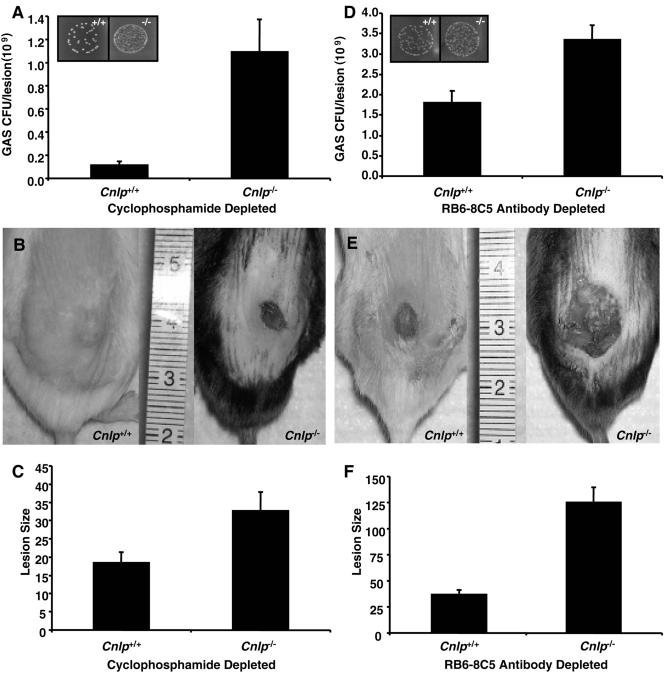
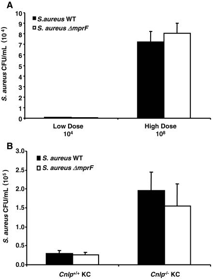
Immune defense at an interface with the external environment reflects the functions of physical and chemical barriers provided by epithelial and immune cells. Resident epithelial cells, such as keratinocytes, produce numerous peptides with direct antimicrobial activity but also provide a physical barrier against invading pathogens and signal the recruitment of circulating immune cells, such as neutrophils. Antimicrobial peptides such as cathelicidin are produced constitutively by neutrophils and are inducible in keratinocytes in response to infection. The multiplicity of antimicrobial peptides and their cellular sources has resulted in an incomplete understanding of the role of cathelicidin production by epithelial cells in cutaneous immune defense. Therefore, this study sought to evaluate keratinocyte antimicrobial activity and the potential contribution of keratinocyte cathelicidin to host protection against two leading human skin pathogens. Wild-type mice and those with a targeted deletion of the cathelicidin gene, Cnlp, were rendered neutropenic prior to cutaneous infection. Interestingly, Cnlp-deficient mice remained more susceptible to group A streptococcus infection than mice with Cnlp intact, suggesting the involvement of epithelial cell-derived cathelicidin in host immune defense. Keratinocytes were then isolated in culture and found to inhibit the growth of Staphylococcus aureus, an effect that was partially dependent on their ability to synthesize and activate cathelicidin. Further, lentivirus-mediated delivery of activated human cathelicidin enhanced keratinocyte antimicrobial activity. Combined, these data illustrate the potential contribution of keratinocyte cathelicidin to the innate immune defense of skin against bacterial pathogens and highlight the need to consider epithelial antimicrobial function in the diagnosis and therapy of skin infection.


Figures

FIG. 1.
FIG. 1.
Neutrophil depletion increases susceptibility to GAS skin infection. (A) H&E staining of excised skin lesions from representative control and depleted Cnlp+/+ mice infected with subcutaneous GAS (inoculum, 4.5 × 10⁵ CFU). Arrows indicate infiltrating leukocytes. Bars, 50 μm. (B) Bacterial counts from excised skin lesions of control and depleted Cnlp+/+ mice infected with GAS (n = 3). Skin lesions were biopsied, homogenized, and plated on TSA to recover GAS.
FIG. 2.
FIG. 2.
Epidermal cathelicidin confers resistance to GAS skin infection in neutropenic mice. (A) Bacterial counts from excised skin lesions of cyclophosphamide-treated Cnlp+/+ and Cnlp/ mice (n = 13) infected with GAS. Mice were administered 250 mg/kg cyclophosphamide i.p. and infected 24 h postdepletion with subcutaneous GAS (inoculum, 4.5 × 10⁵ CFU). Depletion was confirmed by hematology analysis. Inset depicts GAS CFU recovered from skin biopsy specimens on day 7 postinfection. (B) Digital photographs of skin lesions from representative cyclophosphamide-treated Cnlp+/+ and Cnlp/ mice. (C) Sizes of lesions measured from digital photographs of cyclophosphamide-treated mice Cnlp+/+ and Cnlp/ by using NIH Imager. (D) Bacterial counts from excised skin lesions of antibody RB6-8C5-treated Cnlp+/+ and Cnlp/ mice (n = 9) infected with GAS. Mice were administered 450 μg antibody RB6-8C5 i.p. and were infected subcutaneously with GAS (inoculum, 1.5 × 10⁵ CFU) 24 h postdepletion. Depletion was confirmed by hematology analysis. Inset depicts GAS CFU recovered on day 3 postinfection. (E) Digital photographs of skin lesions from representative antibody RB6-8C5-treated Cnlp+/+ and Cnlp/ mice. (F) Sizes of lesions measured from digital photographs of antibody RB6-8C5-treated Cnlp+/+ and Cnlp/ mice by using NIH Imager. All data are means ± standard errors from at least three independent experiments. Statistical analysis was performed using the paired Student t test; P < 0.01 for each set of experiments.
FIG. 3.
FIG. 3.
Keratinocytes demonstrate antimicrobial activity against S. aureus that is dependent on proteolytic activation. (A) Bacterial recovery at 2 h following incubation of log-phase S. aureus ΔmprF (inoculum, 104 CFU/ml) with HaCaT cells overexpressing either hCAP18, cathelin, LL-37, or LacZ. Asterisks, P < 0.001 in comparison with hCAP18. (B) Zones of inhibition (ZOI) measured from radial diffusion assays against S. aureus ΔmprF. Isolated human KC were treated in the presence or absence of PI for 24 h prior to extraction. ZOI were measured using NIH Imager. P < 0.01 for KC versus KC plus PI. (C) Bacterial recovery at 3, 4, and 5 h following incubation of log-phase S. aureus ΔmprF (inoculum, 104 CFU/ml) with isolated human KC treated in the presence or absence of PI for 24 h. “No KC” control wells were incubated with bacteria grown under identical conditions. P < 0.001 for KC versus No KC at 3, 4, and 5 h; P < 0.02 for KC versus KC plus PI at 5 h. All data are means ± standard errors from at least three independent experiments performed in triplicate. Statistical analysis was performed using the paired Student t test, and P values are given for each set of experiments.
FIG. 4.
FIG. 4.
Keratinocytes demonstrate antimicrobial activity against S. aureus that is partially dependent on cathelicidin expression. Bacterial recovery at 3, 4, and 5 h following incubation of log-phase S. aureus ΔmprF (inoculum, 104 CFU/ml) with isolated Cnlp+/+ and Cnlp/ mouse KC is shown. “No KC” control wells were incubated with bacteria grown under identical conditions. Data are means ± standard errors from at least three independent experiments performed in triplicate. Statistical analysis was performed using the paired Student t test. P < 0.001 for Cnlp+/+ KC versus No KC at 3, 4, and 5 h; P < 0.02 for Cnlp+/+ KC versus Cnlp/ KC at 3, 4, and 5 h.
FIG. 5.
FIG. 5.
Keratinocytes internalize S. aureus and demonstrate cathelicidin-dependent intracellular antimicrobial activity. (A) Intracellular bacterial recovery at 2 h following incubation of log-phase S. aureus WT or ΔmprF (low-dose inoculum, 104 CFU; high-dose inoculum, 108 CFU) with isolated human KC. KC were washed and treated with 100 μg/ml gentamicin for 1 h to kill extracellular and cell surface-bound bacteria prior to quantification of intracellular bacteria. (B) Intracellular bacterial recovery at 2 h following incubation of log-phase S. aureus WT or ΔmprF (inoculum, 108 CFU) with isolated Cnlp+/+ and Cnlp/ mouse KC. KC were washed and treated with 100 μg/ml gentamicin for 1 h to kill extracellular and cell surface-bound bacteria prior to quantification of intracellular bacteria. Data are means ± standard errors from three independent experiments performed in triplicate. Statistical analysis was performed using the paired Student t test. P < 0.01 for Cnlp+/+ KC versus Cnlp/ KC with WT S. aureus; P < 0.04 for Cnlp+/+ KC versus Cnlp/ KC with S. aureus ΔmprF.

References

    1. Agerberth, B., J. Charo, J. Werr, B. Olsson, F. Idali, L. Lindbom, R. Kiessling, H. Jornvall, H. Wigzell, and G. H. Gudmundsson. 2000. The human antimicrobial and chemotactic peptides LL-37 and alpha-defensins are expressed by specific lymphocyte and monocyte populations. Blood 96:3086-3093. - PubMed
    1. Anderson, C. 1985. The effect of selected immunomodulating agents on experimental contact reactions. Acta Derm. Venereol. Suppl. (Stockholm) 116:1-48. - PubMed
    1. Bals, R. 2000. Epithelial antimicrobial peptides in host defense against infection. Respir. Res. 1:141-150. - PMC - PubMed
    1. Bals, R., M. J. Goldman, and J. M. Wilson. 1998. Mouse beta-defensin 1 is a salt-sensitive antimicrobial peptide present in epithelia of the lung and urogenital tract. Infect. Immun. 66:1225-1232. - PMC - PubMed
    1. Bals, R., X. Wang, Z. Wu, T. Freeman, V. Bafna, M. Zasloff, and J. M. Wilson. 1998. Human beta-defensin 2 is a salt-sensitive peptide antibiotic expressed in human lung. J. Clin. Investig. 102:874-880. - PMC - PubMed

Publication types

Substances