. 2005 Oct 19;24(20):3543-54.
doi: 10.1038/sj.emboj.7600823. Epub 2005 Sep 22.

Akt phosphorylates acinus and inhibits its proteolytic cleavage, preventing chromatin condensation

Affiliations

Akt phosphorylates acinus and inhibits its proteolytic cleavage, preventing chromatin condensation

Yuanxin Hu et al. EMBO J. .

Abstract

Akt promotes cell survival by phosphorylating and inhibiting components of the intrinsic cell death machinery. Akt translocates into the nucleus upon exposure of cells to survival factors, but little is known about its functions in the nucleus. Here, we show that acinus, a nuclear factor required for apoptotic chromatin condensation, is a direct target of Akt. We demonstrate that Akt phosphorylation of acinus on serine 422 and 573 results in its resistance to caspase cleavage in the nucleus and the inhibition of acinus-dependent chromatin condensation. Abolishing acinus phosphorylation by Akt through mutagenesis accelerates its proteolytic degradation and chromatin condensation. Acinus S422, 573D, a mutant mimicking phosphorylation, resists against apoptotic cleavage and prevents chromatin condensation. Knocking down of acinus substantially decreases chromatin condensation, and depletion of Akt provokes the apoptotic cleavage of acinus. Thus, Akt inhibits chromatin condensation during apoptosis by phosphorylating acinus in the nucleus, revealing a specific mechanism by which nuclear Akt promotes cell survival.


Figures
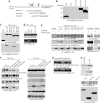
Figure 1
Figure 1
Acinus is a physiological nuclear substrate of Akt. (A) Diagram of acinus-S. Acinus-S possesses three putative Akt phosphorylation motifs (RXRXXS/T) as indicated (▾). The three fragments with each containing a putative phosphorylation motif are indicated with residue numbers. (B) In vitro Akt kinase assay. Purified recombinant GST-fusion proteins were incubated with active Akt. Both fragments B and C were robustly phosphorylated, while fragment A was not. (C) S422 and 573 residues in acinus-S are phosphorylated by Akt. Wild-type fragments, but not S422A and S573A mutants, were phosphorylated (lower panel). Equal amount of GST proteins was employed (upper panel). (D) Phospho-S422 antibody selectively recognizes phosphorylated acinus. While S422 site was markedly phosphorylated in wild-type and S573A acinus, no S422 phosphorylation was detected in S422A and S422, 573A mutants (lower panel). (E) Acinus-S can be phosphorylated in intact cells. HEK 293 cells were transfected with GST–acinus wild-type and mutants. One group was treated with LY294002, and the other group was stimulated by EGF for 20 min. While S422 was markedly phosphorylated in wild-type and S573A acinus upon EGF treatment, no S422 phosphorylation was detected in S422A and S422, 573A mutants (middle left panel). Equal amount of GST proteins was precipitated (top left panel). Akt phosphorylation was also monitored (bottom left panel). Cytosolic and nuclear markers and Akt distribution in the assays (right panels). (F) Endogenous acinus-S can be phosphorylated by Akt in PC12 cells. PC12 cells were pretreated with wortmannin (20 nM) or LY294002 (10 μM) or MEK1 inhibitor PD98059 (10 μM) for 30 min, before NGF was introduced. NGF elicited robust phosphorylation on acinus-S, but PI3K inhibitors markedly blocked it. By contrast, MEK1 inhibitor had no effect (top and second panels). Equal amount of acinus-S was pulled down (third panel). Akt activation was selectively inhibited by PI3K inhibitor, but not MEK1 inhibitor (fourth panel). Cytosolic and nuclear markers and Akt distribution in the assays (right panels). (G) Acinus phosphorylation on acinus (S422A) mutant. Acinus S422A stable cells were induced and treated as described above. PI3K inhibitors substantially blocked NGF-provoked acinus (S573) phosphorylation (upper panel). The S422 phosphorylation was also assessed in NLS-Akt-CA stably transfected PC12 cells. S422 phosphorylation was evident even in control cells, but it was partially blocked by wortmannin (lower panel). (H) Knocking down of Akt blocked acinus S422 phosphorylation (top panel). C-terminus of acinus prevents endogenous acinus phosphorylation by Akt. GST–acinus fragments were transfected into HEK 293 cells, and treated with EGF. Endogenous acinus was analyzed with anti-phospho-422 antibody (middle panel). The expression of transfected GST–acinus fragments (bottom panel).
Figure 2
Figure 2
Akt phosphorylation prevents in vitro acinus proteolytic cleavage. (A) Diagram of acinus-S with putative caspase-3 cleavage sites. Residues 228–335 correspond to p17 active form in acinus-L (aa 987–1093). Caspase cleavage sites and the corresponding fragments with molecular weights were labeled. (B) Akt-phosphorylated fragments resist against apoptotic cleavage. Wild-type phosphorylated proteins resisted against apoptotic degradation, but S422A and S573A mutants were robustly cleaved in apoptotic solution (lower left panel). Equal amounts of GST proteins were employed (upper left panel). Caspase-3 was activated in the cell-free apoptotic solution (right panel). (C) Apoptotic cleavage assay with bacterial expressed full-length GST–acinus-S. Purified GST–acinus proteins were analyzed as described above. Immunoblotting was conducted with anti-acinus antibody after in vitro apoptotic degradation assay. Full-length wild type resisted against caspase cleavage, while S422A and S573A mutants were substantially degraded. Interestingly, S422, 573A mutant was almost completely cleaved. A p30 form of acinus was detected in all samples with highest amount in S422, 573A mutant (lower panel). Equal amount of GST proteins was employed (upper panel). (D) Apoptotic analysis with in vitro transcripted and translated acinus-S. Full-length acinus-S wild type and mutants were labeled with 35S-methionine in rabbit reticulocyte, and incubated with active Akt, then the reaction mixture was assayed in cell-free apoptotic solution. Prominent degradation was observed in both S422A and S422A, 573A mutants, but not in wild-type or S573A sample (bottom panel). Equal amount of 35S-labeled proteins was employed (top panel). S422 was robustly phosphorylated in both wild-type and S573A acinus (middle panel). (E) Apoptotic analysis with in vitro transcripted and translated acinus-S in the absence of Akt. Both S422D and S422, 573D mutants, but not wild-type or S573D sample, resisted against apoptotic degradation.
Figure 3
Figure 3
Akt phosphorylation prevents in vivo acinus proteolytic cleavage. (A) S422A and S422, 573A mutants generate p17 active form in transfected cells. GST–acinus wild-type and mutants were transfected into HEK 293 cells, and treated with 100 μM etoposide for 2 h and followed by 1 μM staurosporine for 7 h. Eto/STS treatment substantially decreased wild-type acinus phosphorylation. As expected, no S422 phosphorylation was detected in S422A or S422, 573A mutant (top panel). Both p45 and p30 bands were produced in acinus-S mutant-transfected cells. S422, 573A exhibited the most abundant p30 form, while no p30 was detected in wild-type transfected cells before apoptotic stimulation. Remarkably, the active p17 form was selectively generated in S422A and S422, 573A mutant cells, but not in wild-type or S573A mutant cells (middle panels). Equal amount of cell lysate was employed (bottom panel). (B) Active nuclear Akt phosphorylates acinus-S and prevents its apoptotic cleavage. PC12 cells were stably transfected with inducible form of Myc-NLS-Akt. Upon induction, cells were subjected to drug treatment. Eto/STS treatment substantially decreased the robust acinus-S S422 phosphorylation in CA cells. By contrast, the faint acinus-S phosphorylation was completely eliminated in both KD and EV cells (top panel). Both p45 and p30 bands were produced in all cells. However, the active p17 form was selectively appeared in KD and EV cells, but not in CA cells (second and third panels). Equal amount of cell lysates was employed (bottom panel). (C) A pan-caspase inhibitor z-VAD-fmk prevents acinus degradation. NLS-Akt stably transfected PC12 cells were pretreated with a pan-caspase inhibitor z-VAD-fmk (20 μM) for 30 min, and followed by the drug treatment. Acinus apoptotic cleavage was almost completely blocked. (D) Depletion of Akt enhances acinus wild-type apoptotic cleavage. PC12 cells were stably transfected with inducible form of Myc-tagged acinus wild-type and S422A. Cells were induced and infected with adenovirus expressing shRNAi of rat Akt1, and pretreated with 50 ng/ml NGF for 1 h, followed by Eto/STS treatment. Knocking down of Akt elicited wild-type acinus cleavage as S422A mutant (upper panel). Endogenous Akt was markedly diminished by its RNAi (lower panel). (E) Inhibition of Akt by wortmannin selectively triggers acinus cleavage. A variety of pharmacological agents were incubated with PC12 cells for 24 h, and the cell lysate was analyzed by immunoblotting with anti-acinus and anti-phospho-Akt antibodies. A phosphoinositol ether analog, a putative Akt inhibitor, fails to incur p30 formation, whereas wortmannin potently provokes acinus degradation. Akt activation was blocked by wortmannin but not by other agents. (F) Acinus stable cells were infected with Akt1 shRNAi for 36 h, then treated with Eto/STS. Depletion of Akt in acinus (S422A), (S573A) and (S422, 573A) cells elicits stronger chromatin condensation than wild-type cells. Chromatin condensation in (S422, 573D) cells is significantly less than that in (S422D) and (S573D) cells.
Figure 4
Figure 4
Akt binds to acinus. (A) Akt co-precipitates with GST–acinus-S. HEK 293 cells were cotransfected with RFP-Akt and GST–acinus-S wild type and mutants, followed by EGF stimulation. Akt strongly binds to acinus-S under basal condition. EGF enhanced the interaction. No significant interaction was observed between acinus mutants and Akt. Interestingly, S422D acinus binds to Akt, which was not regulated by EGF (top panel). S422 was potently phosphorylated in both wild-type and acinus (S573A) mutant (second panel). Equal amount of RFP-Akt and GST–acinus-S was employed (third and bottom panels). (B) Endogenous Akt binds to acinus-S. PC12 cells were respectively pretreated with various inhibitors for 30 min, then NGF was introduced. NGF elicited robust interaction between Akt and acinus-S, but PI3K inhibitors markedly blocked it. By contrast, MEK1 inhibitor had no effect (top panel). Equal amount of Akt was pulled down (middle panel). Equal level of acinus-S was employed (bottom panel). (C) Akt colocalizes with Acinus in the nucleus. HEK 293 cells were cotransfected with RFP-Akt and GST–acinus-S. The transfected cells were stained with anti-GST-FITC antibody. RFP-Akt colocalizes with GST–acinus in the nucleus (left three panels). RFP-Akt alone occurred in both the cytoplasm and the nucleus (right panels). (D) Akt and acinus-S colocalize in the nucleus with speckles. GST–acinus-transfected cells were stained with FITC-conjugated anti-GST antibody and anti-SC35, a specific marker for nuclear speckle.
Figure 5
Figure 5
Nuclear Akt regulates chromatin condensation and DNA fragmentation. (A) Chromatin condensation assay with DAPI. Myc-NLS-Akt-transfected PC12 cells were induced and stimulated with NGF for 30 min, then treated with Eto/STS. The nuclei with condensed chromatin are indicated by white arrowheads (left panels). Quantitative analysis of chromatin condensation (right panel). Numbers of nuclei with condensed chromatin morphology were calculated as means (±s.d.) of five determinations and are representative of three experiments. (B) Characterization of induced nuclear Akt cell lines. Robust induction of CA and KD Akt was detected in the corresponding cell lines (upper panel). In vitro Akt kinase assay demonstrated that Akt in CA cells possesses much higher kinase activity than those in EV and KD cells (lower panel). (C) DNA fragmentation and caspase-3 cleavage. Drug treatment triggered evident DNA fragmentation in KD cells, while less extensive DNA fragmentation was also observed in EV cells. By contrast, no significant DNA fragmentation occurred in CA cells (upper panel). Immunoblotting of caspase-3 (lower panel). (D) Nuclear Akt prevents chromatin condensation and acinus cleavage. PC12 cells were infected with control adenovirus and adenovirus expressing Myr-Akt and NLS-Akt, followed by Eto/STS treatment. NLS-Akt substantially suppressed acinus apoptotic cleavage, but the cytoplasmic Akt failed (top panel). The expression of cytoplasmic and nuclear Akt was verified (middle panel). Compared with control and Myr-Akt-infected cells, chromatin condensation was decreased in NLS-Akt cells (bottom panel).
Figure 6
Figure 6
Acinus-mediated chromatin condensation is Akt-phosphorylation dependent. (A) Acinus cleavage in stably transfected PC12 cell lines. Myc-acinus stably transfected PC12 cells were induced and pretreated with NGF, followed by Eto/STS treatment. The p30 form was evident in all cells, but it was increased after stimulation. P45 form was substantially enhanced after drug treatment. Notably, p17 was selectively generated in S422A or S422, 573A cells after apoptotic stimulation (top panel). (B) Chromatin condensation analysis. The nuclei with condensed chromatin were labeled with white arrowhead (upper panel). About 50% nuclei in S422A and S422, 573A cells displayed condense chromatin morphology, with less than 10% in S573A and wild-type acinus-transfected cells (lower panel). (C) S422, 573D cells resist caspase-induced degradation and chromatin condensation. Stably transfected PC12 cells were induced and followed by Eto/STS treatment in the absence of NGF. Acinus apoptotic cleavage and p17 were substantially decreased in S422D and S422, 573D cells compared with other cell lines (top and middle panels). Chromatin condensation correlated with acinus degradation (lower panel). (D) NGF withdrawal provokes acinus cleavage and chromatin condensation. Stably transfected PC12 cells were incubated with NGF for 5 days. The differentiated cells were cultured in medium without NGF. In 24 h, markedly cleaved p45 and p30 fragments of acinus occurred in S422A and S422AS573A cells, and S573A cells revealed less extent of p45 and p30 fragments. (E) Induction of S422A in PC12 cells does not affect caspase-3 activation. PC12 cells were stably transfected with inducible form of Myc-tagged acinus (S422A). Cells were induced. Immunoblotting analysis was conducted with anti-caspase-3 antibody.
Figure 7
Figure 7
Knocking down of acinus decreases chromatin condensation. (A) Endogenous acinus was decreased upon shRNAi of acinus treatment but not in the control sample, while tubulin control is not affected. (B) NLS-Akt cells were infected with control adenovirus and adenovirus expressing shRNAi to knock down acinus. The infected cells were then treated with Eto/STS. Depletion of acinus abolished p17 formation (left panel). Compared with control cells, depletion of acinus decreased chromatin condensation (right panel).

Similar articles

Cited by

References

    1. Ahmed NN, Franke TF, Bellacosa A, Datta K, Gonzalez-Portal ME, Taguchi T, Testa JR, Tsichlis PN (1993) The proteins encoded by c-akt and v-akt differ in post-translational modification, subcellular localization and oncogenic potential. Oncogene 8: 1957–1963 - PubMed
    1. Ahn JY, Rong R, Liu X, Ye K (2004) PIKE/nuclear PI 3-kinase signaling mediates the antiapoptotic actions of NGF in the nucleus. EMBO J 23: 3995–4006 - PMC - PubMed
    1. Biggs WH III, Meisenhelder J, Hunter T, Cavenee WK, Arden KC (1999) Protein kinase B/Akt-mediated phosphorylation promotes nuclear exclusion of the winged helix transcription factor FKHR1. Proc Natl Acad Sci USA 96: 7421–7426 - PMC - PubMed
    1. Brunet A, Bonni A, Zigmond MJ, Lin MZ, Juo P, Hu LS, Anderson MJ, Arden KC, Blenis J, Greenberg ME (1999) Akt promotes cell survival by phosphorylating and inhibiting a Forkhead transcription factor. Cell 96: 857–868 - PubMed
    1. Brunet A, Datta SR, Greenberg ME (2001) Transcription-dependent and -independent control of neuronal survival by the PI3K–Akt signaling pathway. Curr Opin Neurobiol 11: 297–305 - PubMed

Publication types