. 2005 Oct 19;24(20):3565-75.
doi: 10.1038/sj.emboj.7600820. Epub 2005 Sep 22.

Mediation of Epstein-Barr virus EBNA-LP transcriptional coactivation by Sp100

Affiliations

Mediation of Epstein-Barr virus EBNA-LP transcriptional coactivation by Sp100

Paul D Ling et al. EMBO J. .

Abstract

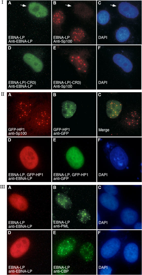
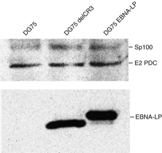
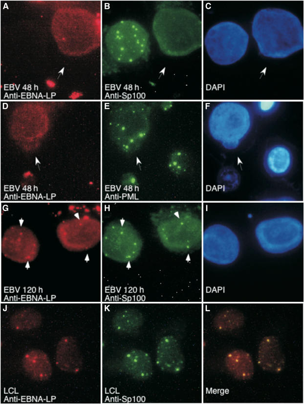
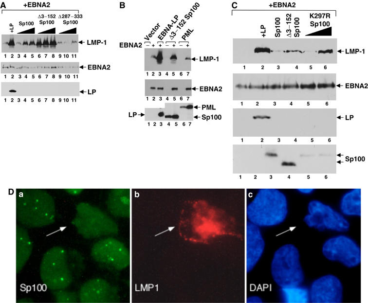
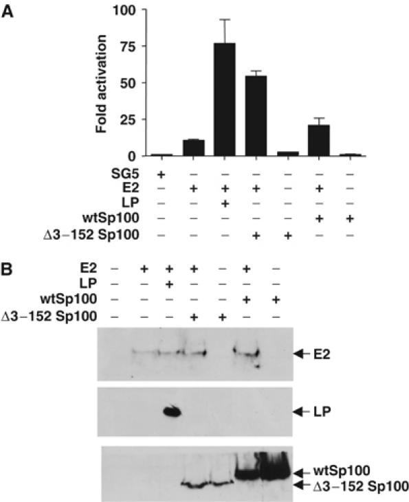
The Epstein-Barr virus (EBV) EBNA-LP protein is important for EBV-mediated B-cell immortalization and is a potent gene-specific coactivator of the viral transcriptional activator, EBNA2. The mechanism(s) by which EBNA-LP functions as a coactivator remains an important question in the biology of EBV-induced B-cell immortalization. In this study, we found that EBNA-LP interacts with the promyelocytic leukemia nuclear body (PML NB)-associated protein Sp100 and displaces Sp100 and heterochromatin protein 1alpha (HP1alpha) from PML NBs. Interaction between EBNA-LP and Sp100 was mediated through conserved region 3 in EBNA-LP and the PML NB targeting domain in Sp100. Overexpression of Sp100 lacking the N-terminal PML NB targeting domain, but not a mutant form of Sp100 lacking the HP1alpha interaction domain, was sufficient to coactivate EBNA2 in a gene-specific manner independent of EBNA-LP. These findings suggest that Sp100 is a major mediator of EBNA-LP coactivation. These studies indicate that modulation of PML NB-associated proteins may be important for establishment of latent viral infections, and also identify a convenient model system to investigate the functions of Sp100.


Figures

Figure 1
Figure 1
Panel I: EBNA-LP, but not EBNA-LP ΔCR3, displaces Sp100 from PML NBs. Expression of EBNA-LP (green, A) in Hep-2 cells resulted in localization of the protein in a diffuse nuclear staining pattern. EBNA-LP displaced Sp100 (red, B) from PML NBs. In contrast, expression of a mutant EBNA-LP, which lacked the CR3 domain (green, D), did not alter the cellular location of Sp100 (red, E). The white arrow in (A–C) points to the EBNA-LP-expressing cell. DAPI staining indicates the location of cell nuclei in (C) and (F). Panel II: EBNA-LP displaces GFP–HP1α from PML NBs. After transfection of Hep-2 cells with a plasmid encoding GFP–HP1α, staining for Sp100 (red, A) and GFP–HP1α (green, B) revealed localization of GFP–HP1α in PML NBs (overlap is shown in yellow in panel C). After expression of EBNA-LP (red, D) and GFP–HP1α in Hep-2 cells, GFP–HP1α did not localize to PML NBs, but was instead distributed diffusely throughout the nucleus (green, E). DAPI staining indicates the location of cell nucleus in (F). Panel III: EBNA-LP did not alter the NB location of PML or CBP. EBNA-LP expression in Hep-2 cells (red, A and D) did not alter the PML NB location of endogenous PML (green B) and CBP (green, E). DAPI staining indicates the location of cell nuclei in (C) and (F).
Figure 2
Figure 2
Immunoblot of DG75 B cells and DG75 B cells constitutively expressing EBNA-LP or ΔCR3LP (DG75 delCR3). Antibodies in serum from PBC patient K142 reacted with Sp100 and PBC autoantigen E2 pyruvate dehydrogenase complex (E2 PDC). Mouse anti-EBNA-LP was used to detect EBNA-LP and ΔCR3LP. There was no difference in the level of Sp100 in the three cell lines.
Figure 3
Figure 3
(A) EBNA-LP interacts with Sp100. DG75 cells cotransfected with Sp100-HA and LP-Flag, or Sp100-HA and ΔCR3LP-Flag, or Sp100-HA and mutCR1aLP-Flag were lysed and precipitated with anti-HA or anti-Flag antibodies. The extracts were divided into equal parts, resolved by SDS–PAGE, and the proteins were detected by immunoblotting with anti-HA (top panel) or anti-EBNA-LP (bottom panel) antibodies. The migration of Sp100, EBNA-LP, and immunoglobulin heavy (Ig H) chain from the primary antibody used in the IP is indicated. As a control, each extract was also treated with Staph A beads alone (no Ab). The coexpressed proteins contained in each extract are indicated above the panel. Precipitation with anti-HA or anti-Flag antibody is indicated above each lane. (B) Schematic of Sp100. Functional domains in Sp100 include amino-acid residues 1–152 (PML NB targeting domain and Sp100 homodimerization region), 287–333 (HP1α interaction domain and SUMO modification site), and 444–450 (nuclear localization sequence). Five Sp100 deletion mutants were used in coimmunoprecipitation and functional studies as indicated. (C) EBNA-LP interacts with the PML NB-targeting domain in Sp100. Lysates from cells cotransfected with EBNA-LP-Flag and one of each of the Sp100-HA deletion mutants were immunoprecipitated with anti-HA (H) or anti-Flag (F) antibodies and the resulting precipitates were probed for Sp100 using anti-HA antibodies. The Δ153–286 Sp100 mutant migrated just below the Ig H chain and is designated by asterisks. Each cell extract was also mock-precipitated as a control (no Ab).
Figure 4
Figure 4
Sp100, but not PML, is displaced from PML NBs following EBV infection of B lymphocytes. At 48 h after EBV infection, Sp100 (green, B) was displaced from PML NBs in nearly all infected cells. EBV-infected cells were identified by staining with anti-EBNA-LP antibodies (red, A, D, G, J). PML (green, E) was not displaced from NBs during EBV infection. At 120 h after infection, both Sp100 (green, H) and EBNA-LP localized to PML NBs. In LCLs, both Sp100 (green, K) and EBNA-LP (red, J) localized to NBs. Overlap between Sp100 and EBNA-LP appears yellow in (L). White arrows in (A–F) indicate EBV-infected cells. White arrows in (G, H) point to examples of EBNA-LP and Sp100 in NBs. DAPI staining indicates the location of nuclei in (C, F, I).
Figure 5
Figure 5
Overexpression of wtSp100 or Δ3–152 Sp100 coactivates EBNA2 in the absence of EBNA-LP. Coactivation of EBNA2 depends on the Sp100–HP1α interaction domain. (A) Eli-BL cells were transfected with EBNA2 or EBNA2 and EBNA-LP or EBNA2 and Sp100. Increasing doses of the Sp100 expression plasmids were cotransfected with EBNA2 as indicated. Cell extracts were probed by Western blot for LMP-1 expression (top panel). Levels of EBNA2 and EBNA-LP were detected in these extracts by Western blot using anti-EBNA2 and anti-EBNA-LP antibodies, respectively. (B) To examine the ability of another NB component to coactivate LMP-1 expression, PML was cotransfected with EBNA2 and the level of LMP-1 was measured by immunoblot. In the absence of EBNA-LP, PML was unable to enhance EBNA2-induced LMP-1 expression above the level of EBNA2 alone (compare lanes 7 and 2). For comparison, the ability of EBNA-LP and Δ3–152 Sp100 to induce LMP-1 expression is shown (lanes 3 and 5). Successful production of EBNA2, EBNA-LP, Sp100, and PML is demonstrated in the immunoblots below (lower panels). (C) Eli-BL cells were transfected with EBNA2 or EBNA2 and EBNA-LP or EBNA2 and various Sp100 mutants. Increasing amounts of the expression plasmid encoding K297R Sp100 were cotransfected with EBNA2 as indicated. Cell extracts were probed by Western blot for LMP-1 expression (top panel). Levels of EBNA2, EBNA-LP, and overexpressed Sp100 proteins were detected in these extracts by Western blot using anti-EBNA2, anti-EBNA-LP, and anti-HA antibodies, respectively, and are shown in the lower panels. (D) Immunofluorescence of Eli-BL cells cotransfected with EBNA2 and EBNA-LP were stained with anti-Sp100 (a) and anti-LMP-1 (b). Sp100 was displaced from PML NBs in all cells that expressed LMP-1. White arrow points to the transfected cell. DAPI staining indicates the location of nuclei in (c).
Figure 6
Figure 6
EBNA-LP independent coactivation of EBNA2 by Sp100 in EBV-negative cells. (A) DG75 cells were transfected with vector alone (SG5), EBNA2 (E2), EBNA-LP (LP), wtSp100, or an Sp100 deletion mutant (Δ3–152 Sp100) as indicated below the graph. Luciferase activity is shown as fold activation above control. Results are an average of three independent experiments with standard errors. (B) Western blots of cell extracts used for the luciferase assays in (A). The top panel shows extracts from each transfection probed for EBNA2 (E2). The middle and bottom panels are identical blots probed with anti-EBNA-LP and anti-HA antibodies, respectively.

Similar articles

Cited by

References

    1. Adamson AL, Kenney S (2001) Epstein–Barr virus immediate-early protein BZLF1 is SUMO-1 modified and disrupts promyelocytic leukemia bodies. J Virol 75: 2388–2399 - PMC - PubMed
    1. Ahn JH, Hayward GS (1997) The major immediate-early proteins IE1 and IE2 of human cytomegalovirus colocalize with and disrupt PML-associated nuclear bodies at very early times in infected permissive cells. J Virol 71: 4599–4613 - PMC - PubMed
    1. Allan GJ, Inman GJ, Parker BD, Rowe DT, Farrell PJ (1992) Cell growth effects of Epstein–Barr virus leader protein. J Gen Virol 73: 1547–1551 - PubMed
    1. Bell P, Lieberman PM, Maul GG (2000) Lytic but not latent replication of Epstein–Barr virus is associated with PML and induces sequential release of nuclear domain 10 proteins. J Virol 74: 11800–11810 - PMC - PubMed
    1. Bloch DB, Chiche JD, Orth D, de la Monte SM, Rosenzweig A, Bloch KD (1999) Structural and functional heterogeneity of nuclear bodies. Mol Cell Biol 19: 4423–4430 - PMC - PubMed

Publication types

MeSH terms