Comparative Study
. 2005 Oct 4;102(40):14392-7.
doi: 10.1073/pnas.0502896102. Epub 2005 Sep 22.

Rapid directional shift of mitochondrial DNA heteroplasmy in animal tissues by a mitochondrially targeted restriction endonuclease

Affiliations
Comparative Study

Rapid directional shift of mitochondrial DNA heteroplasmy in animal tissues by a mitochondrially targeted restriction endonuclease

Maria Pilar Bayona-Bafaluy et al. Proc Natl Acad Sci U S A. .

Abstract

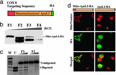
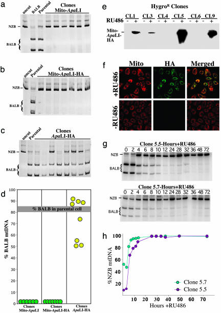
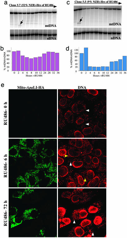
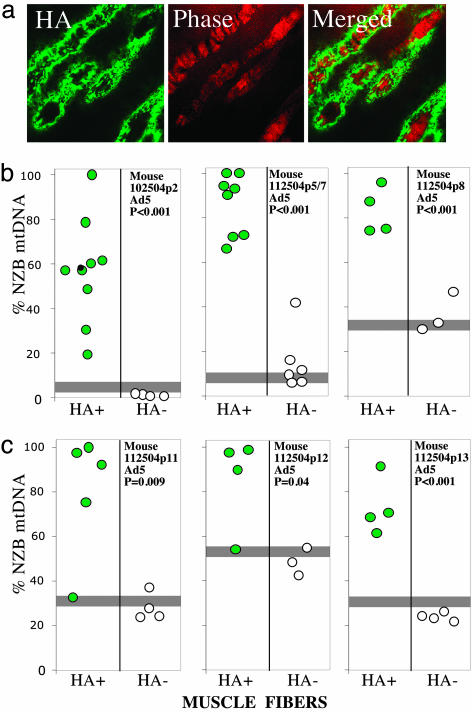
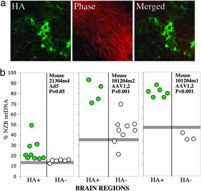
Frequently, mtDNA with pathogenic mutations coexist with wild-type genomes (mtDNA heteroplasmy). Mitochondrial dysfunction and disease ensue only when the proportion of mutated mtDNAs is high, thus a reduction in this proportion should provide an effective therapy for these disorders. We developed a system to decrease specific mtDNA haplotypes by expressing a mitochondrially targeted restriction endonuclease, ApaLI, in cells of heteroplasmic mice. These mice have two mtDNA haplotypes, of which only one contains an ApaLI site. After transfection of cultured hepatocytes with mitochondrially targeted ApaLI, we found a rapid, directional, and complete shift in mtDNA heteroplasmy (2-6 h). We tested the efficacy of this approach in vivo, by using recombinant viral vectors expressing the mitochondrially targeted ApaLI. We observed a significant shift in mtDNA heteroplasmy in muscle and brain transduced with recombinant viruses. This strategy could prevent disease onset or reverse clinical symptoms in patients harboring certain heteroplasmic pathogenic mutations in mtDNA.


Figures

Fig. 1.
Fig. 1.
Construction and characterization of a Mito-ApaLI-HA. (a) The genetic code of a synthetic ApaLI gene was optimized for mammalian translation and a HA-tag coding sequence added to the 3′ end. The gene was cloned downstream of the cytochrome oxidase subunit 8 (COX 8) mitochondrial targeting sequence in a pcDNA3 vector. (b) Mito-ApaLI-HA containing the COX 8 targeting sequence was expressed in human embryonic kidney 293T cells and after 48 h, an S-100 fraction was purified by heparin Sepharose binding. Fractions eluted with increasing salt concentrations (F1-F4) were analyzed by immunoblot using an anti-HA antibody. Fraction 1 shows mostly full length protein, whereas the other lanes have degradation products. (c) Fraction F1 was able to digest a DNA fragment at the ApaLI cleavage sites. Control (C) is the same PCR fragment digested with commercial ApaLI. (d) pcDNA3-based plasmids harboring either Mito-ApaLI-HA or ApaLI-HA constructs were transiently transfected into COS-7 cells. After 24 h, cells were stained with the mitochondrial specific dye Mitotracker red, fixed, and immunostained for the HA epitope. In the merged image, note the mitochondrial colocalization of HA immunofluorescence (in green) and Mitotracker (in red) in cells expressing Mito-ApaLI-HA.
Fig. 2.
Fig. 2.
Rapid and complete shift in mtDNA heteroplasmy by Mito-ApaLI-HA. Cultured hepatocyte clones from heteroplasmic BALB/NZB mice stably transfected with the different ApaLI constructs were analyzed for their mtDNA haplotypes. Cells were collected after selection in G418. DNA was purified and used to amplify a PCR fragment spanning an ApaLI site. After digestion with ApaLI, the samples were analyzed by PAGE (“uncut,” PCR fragment not digested; “BALB,” PCR fragment from a BALB mouse digested with ApaLI; “Parental,” PCR fragment from untransfected hepatocytes digested with ApaLI). a-c show autoradiographies of PAGE/RFLP of amplicons from clones expressing the different ApaLI constructs. Mitochondrially targeted ApaLI promoted a complete shift in mtDNA heteroplasmy toward the NZB haplotype (no ApaLI sites), irrespective of the presence of the HA tag (a and b). c shows that in the absence of a mitochondrial targeting sequence, no mtDNA heteroplasmy shift took place. d summarizes the data from the individual hepatocyte clones. e shows the immunoblot (anti-HA) analysis of clones expressing an inducible form of mito-ApaLI-HA. A strong induction of mito-ApaLI-HA expression can be observed in some hepatocyte clones after the addition of 10 nM RU486. f shows the RU486 induction of Mito-ApaLI-HA by immunocytochemistry in clone 5. g shows the time course of the shift in mtDNA heteroplasmy after induction of Mito-ApaLI-HA expression in clones 5.5 and 5.7. h shows a similar analysis, after digestion of PCR fragments with HindIIII (also discriminates BALB and NZB mtDNA), in graphic format.
Fig. 3.
Fig. 3.
Transient mtDNA depletion associated with expression of Mito-ApaLI-HA. Cells were collected at different time points after expression of Mito-ApaLI-HA, and their DNA was isolated and analyzed by Southern blot. Total DNA was digested with SacI, which cuts both BALB and NZB mtDNA once at nucleotide 9047, probed to a mtDNA probe and subsequently to a nuclear 18SrDNA probe. a and c show the Southern blot, whereas b and d show the quantitation of mtDNA/nuclear DNA ratios. A fragment with the size predicted from a double-digestion SacI+ApaLI was observed only at 2-4 h of Mito-ApaLI-HA induction (arrow). (e) Cells with 9% NZB mtDNA were induced to express Mito-ApaLI-HA with RU486. The cells were fixed at different time points after induction and immunostained for HA (green) and DNA (red). Note that at 6-h induction, cells expressing Mito-ApaLI-HA lost the punctate nucleoid structures (white arrowhead) that were substituted by a diffuse crossreacting material (yellow arrowheads). However, nucleoids were reestablished in Mito-ApaLI-HA-expressing cells at 72-h induction.
Fig. 4.
Fig. 4.
Expression of Mito-ApaLI-HA in muscle leads to efficient shift in mtDNA heteroplasmy. Skeletal muscle (gastrocnemius) of 5-day-old mice was injected with Ad5-Mito-ApaLI-HA, as described in Methods. After 1 (b) or 2 (c) weeks, animals were killed and muscle frozen in liquid nitrogen-cooled isopentane. Twenty-micrometer muscle sections were immunostained for HA (a) and microdissected using laser capture microscopy. Microdissected samples were treated with an alkaline lysis solution and subjected to PCR/RFLP for mtDNA haplotype determination. The proportions of NZB/BALB mtDNA were calculated after “last cycle hot” PCR, digestion with ApaLI, PAGE, and phosphorimaging (b and c). Horizontal gray bars represent the percent NZB mtDNA in the mouse tail.
Fig. 5.
Fig. 5.
Expression of Mito-ApaLI-HA in brain leads to a shift in mtDNA heteroplasmy. The putamen of anesthetized 2- to 4-mo-old mice was stereotactically injected with 2 μl of virus suspensions (Ad5-Mito-ApaLI-HA or AAV1,2-Mito-ApaLI-HA). After 1 (Ad5) or 2 weeks (AAV1,2), the animals were killed and the brains snap-frozen in liquid nitrogen. Twenty-micrometer sections were screened for GFP expression (not shown) and adjacent sections stained for HA expression (a). Regions of positive and negative staining were microdissected by laser capture microscopy and subjected to mtDNA haplotype analysis as described in the legend to Fig. 4b. Horizontal gray bars represent the percent NZB mtDNA in the mouse tail.

References

    1. Kaufman, B. A., Newman, S. M., Hallberg, R. L., Slaughter, C. A., Perlman, P. S. & Butow, R. A. (2000) Proc. Natl. Acad. Sci. USA 97, 7772-7777. - PMC - PubMed
    1. Legros, F., Malka, F., Frachon, P., Lombes, A. & Rojo, M. (2004) J. Cell Sci. 117, 2653-2662. - PubMed
    1. Bogenhagen, D. F., Wang, Y., Shen, E. L. & Kobayashi, R. (2003) Mol. Cell Proteomics 2, 1205-1216. - PubMed
    1. Garrido, N., Griparic, L., Jokitalo, E., Wartiovaara, J., van der Bliek, A. M. & Spelbrink, J. N. (2003) Mol. Biol. Cell 14, 1583-1596. - PMC - PubMed
    1. Thorburn, D. R. & Dahl, H. H. (2001) Am. J. Med. Genet. 106, 102-114. - PubMed

Publication types