. 2005 Sep 23;309(5743):2061-4.
doi: 10.1126/science.1118046.

Xanthorhodopsin: a proton pump with a light-harvesting carotenoid antenna

Affiliations

Xanthorhodopsin: a proton pump with a light-harvesting carotenoid antenna

Sergei P Balashov et al. Science. .

Abstract

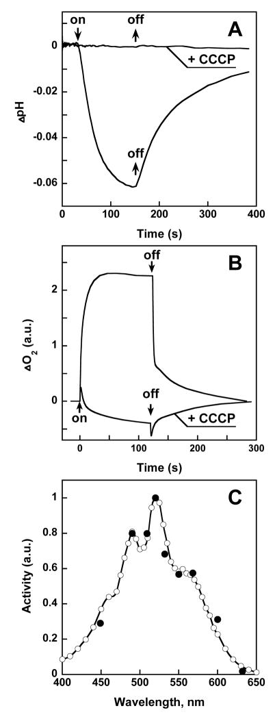
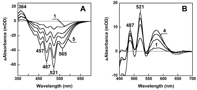
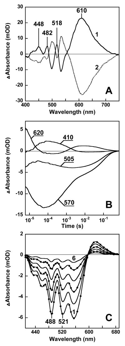
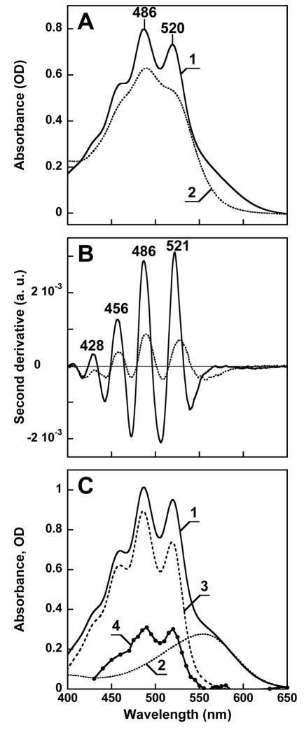
Energy transfer from light-harvesting carotenoids to chlorophyll is common in photosynthesis, but such antenna pigments have not been observed in retinal-based ion pumps and photoreceptors. Here we describe xanthorhodopsin, a proton-pumping retinal protein/carotenoid complex in the eubacterium Salinibacter ruber. The wavelength dependence of the rate of pumping and difference absorption spectra measured under a variety of conditions indicate that this protein contains two chromophores, retinal and the carotenoid salinixanthin, in a molar ratio of about 1:1. The two chromophores interact strongly, and light energy absorbed by the carotenoid is transferred to the retinal with a quantum efficiency of approximately 40%. The antenna carotenoid extends the wavelength range of the collection of light for uphill transmembrane proton transport.


Figures

Fig. 1
Fig. 1
Light-induced proton transport in Salinibacter ruber, and its action spectrum. A. pH change in a membrane vesicle suspension produced by illumination at 550 nm at pH 6.5, in the absence and presence of 20 μM protonophore CCCP (carbonyl cyanide m-chlorophenyl-hydrazone), at a light intensity of 2 mW/cm2. B. Kinetics of light-induced changes in the rate of oxygen uptake caused by photo-inhibition of respiration in the cell suspension of Salinibacter ruber, in the absence and presence of 1 μM CCCP. C. Action spectra for photo-inhibition of the respiration by intact cells (open circles) and light-induced proton transport in membrane vesicles, latter measured as in Fig. 1 (closed circles). In both cases the optical density of the sample was below 0.2.
Fig. 2
Fig. 2
Spectroscopic detection of the retinal protein of Salinibacter. A. Curves 1 through 5, difference spectra upon illumination of a cell membrane suspension at >550 nm in the presence of 0.2 M hydroxylamine for 4, 12, 20, 36, and 60 min at 20°C, respectively. B. Curves 1 through 4, difference spectra measured 5, 15, 25, and 60 min after addition of all-trans retinal to the Salinibacter ruber cell membranes which had been bleached with 0.2 M hydroxylamine.
Fig. 3
Fig. 3
Absorption changes during the photocycle. A. Light-induced absorption changes in water-glycerol suspension of cell membranes at 175 K. Spectrum 1, illumination of membranes with 520 nm light, spectrum 2, subsequent illumination at > 650 nm. B. Kinetics of laser flash induced absorption changes at selected characteristic wavelengths in membranes of Salinibacter solubilized with 0.15% dodecyl maltoside, in 100 mM NaCl, pH 8.8, 20°C. The global fit indicates that kinetics include at least six components, with time-constants of 7.5 μs, 35 μs, 280 μs, 1.3 ms, 11 ms, and 100 ms. C. Transient difference spectra during the photocycle, measured at 10, 30, 60, 100, 160, and 250 ms (curves 1 through 6) after a 532 nm laser flash. Conditions and sample are as in B.
Fig. 4
Fig. 4
Absorption spectrum of xanthorhodopsin: its carotenoid and retinal chromophore components, and estimation of the efficiency of energy transfer from the carotenoid. A. Absorption spectrum of solubilized and purified xanthorhodopsin, before (spectrum 1) and after (spectrum 2) bleaching with hydroxylamine. B. Second derivatives of the two spectra in A. C. Estimated absorption spectrum of the complex and the contribution of carotenoid to it and to the action spectrum. Spectrum 1: calculated spectrum of xanthorhodopsin; spectrum 2: bacteriorhodopsin, 10 nm shifted to the blue and appropriately scaled to model the retinal component of xanthorhodopsin; spectra 3 and 4: carotenoid component of the absorption spectrum and the action spectrum, respectively, obtained by subtracting the contribution of the retinal.

Similar articles

Cited by

References

    1. Antón J, Rosselló-Mora R, Rodríguez-Valera F, Amann R. Appl Environ Microbiol. 2000;66:3052. - PMC - PubMed
    1. Antón J, et al. Int J Syst Evol Micrbiol. 2002;52:485. - PubMed
    1. Oren A, Heldal M, Norland S, Galinski EA. Extremophiles. 2002;6:491. - PubMed
    1. Lutnaes BF, Oren A, Liaaen-Jensen S. J Nat Prod. 2002;65:1340. - PubMed
    1. Oesterhelt D, Stoeckenius W. Nature. 1971;233:149. - PubMed

Publication types

MeSH terms